. 2004 Jun 16;23(12):2381-91.
doi: 10.1038/sj.emboj.7600241. Epub 2004 Jun 3.

A subset of human 35S U5 proteins, including Prp19, function prior to catalytic step 1 of splicing

Affiliations

A subset of human 35S U5 proteins, including Prp19, function prior to catalytic step 1 of splicing

Olga V Makarova et al. EMBO J. .

Abstract

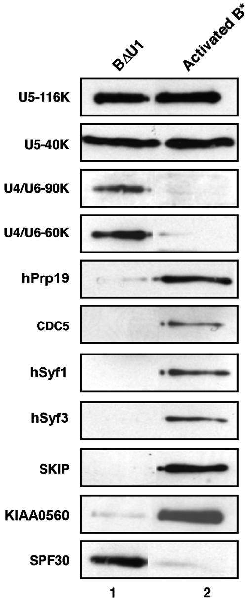
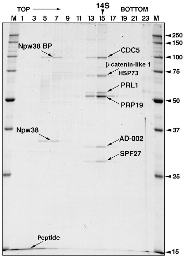
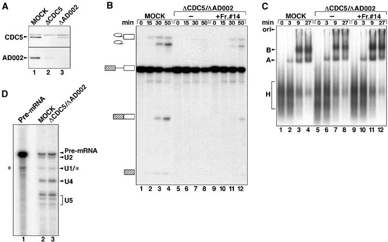
During catalytic activation of the spliceosome, snRNP remodeling events occur, leading to the formation of a 35S U5 snRNP that contains a large group of proteins, including Prp19 and CDC5, not found in 20S U5 snRNPs. To investigate the function of 35S U5 proteins, we immunoaffinity purified human spliceosomes that had not yet undergone catalytic activation (designated BDeltaU1), which contained U2, U4, U5, and U6, but lacked U1 snRNA. Comparison of the protein compositions of BDeltaU1 and activated B* spliceosomes revealed that, whereas U4/U6 snRNP proteins are stably associated with BDeltaU1 spliceosomes, 35S U5-associated proteins (which are present in B*) are largely absent, suggesting that they are dispensable for complex B formation. Indeed, immunodepletion/complementation experiments demonstrated that a subset of 35S U5 proteins including Prp19, which form a stable heteromeric complex, are required prior to catalytic step 1 of splicing, but not for stable integration of U4/U6.U5 tri-snRNPs. Thus, comparison of the proteomes of spliceosomal complexes at defined stages can provide information as to which proteins function as a group at a particular step of splicing.


Figures

Figure 1
Figure 1
Characterization of immunoaffinity-purified BΔU1 spliceosomes. (A) RNA composition of BΔU1 versus activated B* spliceosomes. RNAs extracted from gradient-purified BΔU1 (lane 1), B* (lane 2) or total snRNPs (lane 3) were analyzed by denaturing PAGE and visualized by silver staining. (B) Identification of RNA base-pairing interactions in BΔU1. Gradient-fractionated BΔU1 (lanes 1–2, 5–6, and 11–12), B* (lanes 3–4, 7–8, and 13–14), or U4/U6.U5 tri-snRNPs (lanes 9–10 and 15–16) were incubated in the absence (even lanes) or presence (odd lanes) of psoralen (AMT) prior to UV irradiation. RNA was analyzed by denaturing PAGE, followed by either autoradiography to directly detect the 32P-labeled pre-mRNA present in the BΔU1 and B* spliceosomes (lanes 1–4), or by Northern blotting with 32P-labeled probes specific for U6 (lanes 5–10) or U2 (lanes 11–16). (C) Migration behavior of purified BΔU1 spliceosomes on native agarose gels. 32P-labeled MINX pre-mRNA was incubated under splicing conditions with HeLa nuclear extract for 0–30 min (lanes 1–5), and spliceosome assembly was analyzed on a 2% agarose gel, together with immunoaffinity-purified BΔU1 (lane 6). The positions of the H, A, and B complexes are indicated on the left.
Figure 2
Figure 2
BΔU1 spliceosomes catalyze splicing in the absence of exogenously added snRNPs. Immunoaffinity-purified BΔU1 spliceosomes (SPL) (lanes 1–7) or 32P-labeled MINX pre-mRNA (RNA) (lanes 8–14) were incubated for 0–80 min (as indicated) under splicing conditions in the presence of 20% micrococcal nuclease-treated nuclear extract. RNAs were analyzed by denaturing PAGE and visualized by autoradiography. The positions of the pre-mRNA, and the splicing intermediates and products, are indicated on the left.
Figure 3
Figure 3
Protein composition of BΔU1 versus activated B* spliceosomes. BΔU1 and B* spliceosomes were immunoaffinity purified under identical conditions and subjected to glycerol gradient centrifugation. Proteins from BΔU1 or B* were separated by SDS–PAGE, blotted onto a membrane and immunostained with affinity-purified antibodies against selected proteins, as indicated on the right.
Figure 4
Figure 4
Sedimentation behavior and protein composition of CDC5/Prp19-containing complexes. Complexes were immunoaffinity purified from HeLa nuclear extract with anti-CDC5 antibodies and subjected to 5–20% glycerol gradient centrifugation. Proteins from uneven fractions were separated by SDS–PAGE and stained with Coomassie. The following proteins (indicated with arrows) were identified by MS: CDC5 (NP_001244), HSP73 (NP_006588), β-catenin-like 1 (NP_110517), PRL1 (NP_002660), Prp19 (NP_055317), AD002 (AAF14858), SPF27 (NP_005863), Npw38 (NP_005701), and Npw38BP (NP_057396).
Figure 5
Figure 5
Spliceosomal B complex forms in the absence of the CDC5/PRP19 complex. (A) Nuclear extracts are efficiently immunodepleted of CDC5 and AD002. Western blot of mock-depleted (lane 1), CDC5-depleted (lane 2) or AD002-depleted (lane 3) extract probed with anti-CDC5 (upper panel) or anti-AD002 (lower panel) antibodies. (B) Time course of splicing of [32P] MINX pre-mRNA in mock-depleted extract (MOCK, lanes 1–4) or extract depleted with CDC5 and AD002 (ΔCDC5/ΔAD002, lanes 5–8), or CDC5/AD002-depleted extract complemented with purified CDC5/PRP19 complex (fraction no. 14, from the gradient in Figure 4) (lanes 9–12). RNA was analyzed by denaturing PAGE and visualized by autoradiography. The pre-mRNA, and splicing intermediates and products are indicated on the left. (C) The CDC5/PRP19 complex is not required for B-complex formation. Spliceosome assembly in mock-depleted extract, CDC5/AD002-depleted extract, or CDC5/AD002-depleted extract complemented with the CDC5/PRP19 complex (lanes marked as in (B)) was analyzed at the indicated times by native gel electrophoresis and visualized by autoradiography. The position of the H, A, and B complexes is indicated on the left. (D) Spliceosomes formed in CDC5/AD002-depleted extract contain U4 snRNA. Spliceosomes were immunoprecipitated from mock- (lane 2) or CDC5/AD002-depleted extract (lane 3) with anti-61K antibodies. RNA was isolated, end labeled with 32P-pCp, fractionated by denaturing PAGE, and visualized by autoradiography. The identities of the RNAs are indicated on the right. Internally radiolabeled MINX pre-mRNA (lane 1) was analyzed in parallel. Note that the band migrating at the position of U1 may also contain a pre-mRNA degradation product (*).

References

    1. Ajuh P, Kuster B, Panov K, Zomerdijk JC, Mann M, Lamond AI (2000) Functional analysis of the human CDC5L complex and identification of its components by mass spectrometry. EMBO J 19: 6569–6581 - PMC - PubMed
    1. Ben-Yehuda S, Dix I, Russell CS, McGarvey M, Beggs JD, Kupiec M (2000) Genetic and physical interactions between factors involved in both cell cycle progression and pre-mRNA splicing in Saccharomyces cerevisiae. Genetics 156: 1503–1517 - PMC - PubMed
    1. Brow DA (2002) Allosteric cascade of spliceosome activation. Annu Rev Genet 36: 333–360 - PubMed
    1. Burge CB, Tuschl T, Sharp PA (1999) Splicing of precursors to mRNAs by the spliceosomes. In The RNA World, Gesteland C, Atkins (eds), 2nd edn, Cold Spring Harbor, NY: Cold Spring Harbor Laboratory Press pp 525–560
    1. Chan SP, Kao DI, Tsai WY, Cheng SC (2003) The Prp19p-associated complex in spliceosome activation. Science 302: 279–282 - PubMed

Publication types

LinkOut - more resources