. 2004 Jul 7;23(13):2664-73.
doi: 10.1038/sj.emboj.7600264. Epub 2004 Jun 3.

Positive and negative regulation of SMC-DNA interactions by ATP and accessory proteins

Affiliations

Positive and negative regulation of SMC-DNA interactions by ATP and accessory proteins

Michiko Hirano et al. EMBO J. .

Abstract

Structural maintenance of chromosomes (SMC) proteins are central regulators of higher-order chromosome dynamics from bacteria to humans. The Bacillus subtilis SMC (BsSMC) homodimer adopts a V-shaped structure with an ATP-binding catalytic domain at each end. We report here that two small proteins, ScpA and ScpB, associate with the catalytic domains of BsSMC in an ordered fashion and suppress its ATPase activity. When combined with a 'transition state' mutant of BsSMC that poorly hydrolyzes ATP, ScpA promotes stable engagement of two catalytic domains in an ATP-dependent manner. In solution, this occurs intramolecularly and closes the DNA-entry gate of an SMC dimer. ScpB further stabilizes this conformation and prevents BsSMC from binding to double-stranded DNA (dsDNA). In contrast, when the mutant BsSMC is first allowed to interact with dsDNA, subsequent addition of ScpA leads to assembly of large nucleoprotein complexes, possibly by stabilizing intermolecular engagement of the catalytic domains from different SMC dimers. We propose that the ATP-modulated engagement/disengagement cycle of SMC proteins plays both positive and negative roles in their dynamic interactions with dsDNA.


Figures

Figure 1
Figure 1
Physical interactions among BsSMC, ScpA and ScpB. (A) Purified fractions of BsSMC, ScpA and ScpB were subjected to 10% SDS–PAGE and visualized by staining with Coomassie blue. (B) Two-armed (wild-type) BsSMC was mixed with buffer alone (lanes 1 and 5), ScpA (lanes 2 and 6), ScpB (lanes 3 and 7) or ScpA and ScpB (lanes 4 and 8), and immunoprecipitated with an antibody specific to BsSMC. In all, 10% of input (lanes 1–4) and precipitated fractions (lanes 5–8) were analyzed by immunoblotting with the indicated antibodies. The interaction of ScpA and ScpB with single-armed BsSMC was characterized in the same way (lanes 9–12). (C) Fractions containing ScpA (lanes 1, 4 and 7), ScpB (lanes 2, 5 and 8), or ScpA and ScpB (lanes 3, 6 and 9) were immunoprecipitated with anti-ScpA (lanes 4–6) or anti-ScpB (lanes 7–9). A total of 10% of input (lanes 1–3) and precipitated fractions (lanes 4–9) were analyzed by immunoblotting with the indicated antibodies. (D) ScpA was mixed with two-armed BsSMC (lanes 1 and 6), single-armed BsSMC (lanes 2 and 8), head-less BsSMC (lanes 3 and 10) or hinge-stalk BsSMC (lanes 4 and 12), and immunoprecipitated with anti-ScpA. In all, 10% of input (lanes 1–4) and precipitated fractions (lanes 6, 8, 10 and 12) were analyzed by immunoblotting with the indicated antibodies. To demonstrate the specificity of the immunoprecipitation reactions, ScpA was omitted in lanes 5, 7, 9 and 11.
Figure 2
Figure 2
Reconstitution of a BsSMC–ScpA–ScpB complex in vitro. (A) Purified ScpA (top) or ScpB (bottom) was loaded onto a 5–20% sucrose gradient and centrifuged at 42 000 rpm for 24 h in an SW50.1 rotor. Fractions were subjected to SDS–PAGE and stained with Coomassie blue. (B) Purified BsSMC (top) or a mixture of BsSMC, ScpA and ScpB (bottom) was loaded onto a 5–20% sucrose gradient and centrifuged at 45 000 rpm for 15 h in an SW50.1 rotor. Fractions were analyzed as above. The positions of protein standards (ovalbumin (3.7 S), BSA (4.6 S) and aldolase (7.3 S)) are indicated.
Figure 3
Figure 3
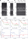
Modulation of SMC activities by ScpA and ScpB. (A) A fixed concentration of dsDNA (pBluescript; 15.6 μM nucleotides) and ssDNA (φ × 174 virion DNA; 15.6 μM nucleotides) was incubated with increasing concentrations of wild-type BsSMC (0, 70, 140, 210, 280, 350 and 420 nM arms) in the absence of ATP (lanes 1–7). The reaction mixtures were fractionated on a 0.7% agarose gel and visualized by EtBr stain. Protein–DNA complexes are indicated by asterisks, and free DNAs are indicated by arrows. (B) dsDNA or ssDNA was incubated with no protein (lane 1), ScpA (840 nM monomers; lane 2), ScpB (840 nM monomers; lane 3) or ScpA and ScpB (840 nM each; lane 4) in the absence of ATP and analyzed as above. (C) dsDNA or ssDNA was incubated with no protein (lane 1), BsSMC alone (lanes 2, 6 and 10), BsSMC and ScpA (lanes 3, 7 and 11), BsSMC and ScpB (lanes 4, 8 and 12) or BsSMC, ScpA and ScpB (lanes 5, 9 and 13) in the absence (lanes 2–5) or presence of ATP (lanes 6–9) or ATPγS (lanes 10–13), and analyzed as above. The final concentrations of BsSMC, ScpA and ScpB used were 420, 840 and 840 nM, respectively. (D) A fixed concentration of wild-type BsSMC (300 nM arms) was mixed with increasing molar ratios of ScpA (red), ScpB (black) or ScpA and ScpB (green). The ATPase activity of the mixtures was assayed in a buffer containing 10 mM KCl and 2 mM MgCl2 in the absence of DNA (panel 1) or in the presence of dsDNA (31.2 μM nucleotides; panel 2) or ssDNA (31.2 μM nucleotides; panel 3). The activity is represented as percentage of the activity of BsSMC without Scp proteins under each condition. Alternatively, the ATPase activity of wild-type BsSMC (300 nM arms) was assayed at different MgCl2 concentrations in the absence (blue) or presence (red) of a fixed molar ratio of ScpA (SMC:ScpA 1:4; panels 4–6). The rate of ATP hydrolysis is expressed as the number of ATP molecules hydrolyzed per second per arm.
Figure 4
Figure 4
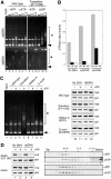
Characterization of the transition state mutant of BsSMC. (A) A fixed concentration of dsDNA or ssDNA (15.6 μM nucleotides) was incubated with no protein (lane 1) or increasing concentrations (105, 210 or 420 nM arms) of the wild-type (lanes 2–7) or E1118Q mutant (lanes 8–13) protein in the absence or presence of ATP. The reaction mixtures were analyzed as in Figure 3A–C. Protein–DNA complexes are indicated by asterisks, and free DNAs are indicated by arrows. (B) The ATPase rates of wild-type BsSMC (WT), transition state mutant (TR) and Walker A mutant (WA) were determined in a buffer containing 5 mM KCl and 2 mM MgCl2 in the absence or presence of dsDNA or ssDNA. The rate of ATP hydrolysis is expressed as the number of ATP molecules hydrolyzed per second per arm. (C) A fixed concentration of wild-type or mutant BsSMC proteins (420 nM arms) was incubated with dsDNA in the absence or presence of ATP. The reaction mixtures were split into two aliquots, and subjected to gel-shift (lanes 2–9) and spin-down (lanes 12 and 13) assays. In the latter, the mixtures were spun at 16 000 g for 15 min, and the supernatant (sup) and pellet (ppt) fractions were analyzed by 7.5% SDS–PAGE. Lane 1 contained no protein, and lanes 10 and 11 contained no dsDNA. (D) The AAAA-E1118Q and AAAA mutants were subjected to the spin-down assay as above in the absence (left, lanes 1 and 2) or presence (left, lanes 3 and 4) of dsDNA. Alternatively, the DNA-free reactions were fractionated by 5–20% sucrose gradients and the fractions were analyzed by SDS–PAGE (right).
Figure 5
Figure 5
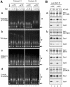
Effects of ScpA and ScpB on BsSMC mutants defective in the ATPase cycle. (A) The dsDNA-binding activities of four different BsSMC proteins (the transition state mutant E1118Q (a), wild-type BsSMC (b), the Walker A mutant K37I (c) and the C-motif mutant S1090R (d)) in the presence or absence of ScpA and/or ScpB were examined by gel-shift assays with two different protocols. In Protocol #1, BsSMC was first incubated with buffer alone (lanes 2 and 6), ScpA (lanes 3 and 7), ScpB (lanes 4 and 8) or ScpA and ScpB (lanes 5 and 9) in the absence or presence of ATP, and then dsDNA was added to the protein mixtures. In Protocol #2, dsDNA was first incubated with BsSMC alone in the absence or presence of ATP, and then supplemented with buffer alone (lanes 10 and 14), ScpA (lanes 11 and 15), ScpB (lanes 12 and 16) or ScpA and ScpB (lanes 13 and 17). The molar ratio of input proteins (SMC:ScpA:ScpB) was 1:2:2. No protein was added in lane 1. After incubation, the reaction mixtures were fractionated on 0.7% agarose gels and visualized by EtBr stain. Protein–DNA complexes are indicated by asterisks and free DNAs are indicated by arrows. (B) Interactions of four different BsSMC proteins with ScpA and ScpB were assayed by immunoprecipitation. BsSMC was incubated with ScpA and ScpB in the absence (lanes 1 and 2) or presence (lanes 3 and 4) of ATP, and immunoprecipitated with anti-BsSMC. Each of the precipitates was split into two aliquots and washed with a buffer containing 0.1 M KCl (lanes 1 and 3) or 0.5 M KCl (lanes 2 and 4) in the continued absence or presence of ATP. Precipitated fractions were analyzed by immunoblotting with the indicated antibodies.
Figure 6
Figure 6
A model for positive and negative regulation of SMC–dsDNA interactions by ATP and accessory proteins. In the absence of dsDNA, BsSMC undergoes a cycle of ATP binding, engagement and ATP hydrolysis (stages 1–3). The Walker A (K37I), C-motif (S1090R) or transition state (E1118Q) mutation specifically blocks each of these steps as indicated. The ATP-dependent engagement of two catalytic domains occurs only intramolecularly (stage 3) unless the hinge structure is disturbed. ScpA and ScpB associate with the catalytic domains of BsSMC in a sequential manner (stages 4 and 5). While this occurs regardless of the nucleotide-binding state of BsSMC, only one pathway starting from stage 3 is shown here for simplicity. Association of ScpA partially blocks the dsDNA-binding activity of BsSMC (stage 4), and further association of ScpB blocks it more severely (stage 5). BsSMC binds to dsDNA in an apparently cooperative manner either in the absence (stage 6) or presence (stage 7) of ATP (basal binding). When ATP-dependent engagement of the two catalytic domains is stabilized by the E1118Q mutation, stable dsDNA binding is observed (stages 8 and 9). Subsequent binding of ScpA to BsSMC further stabilizes the engaged state by inhibiting hydrolysis of the bound ATP (stages 10 and 11). The ATP-dependent engagement could occur either intramolecularly through an ‘embrace' mechanism (stages 8 and 10) or intermolecularly through a ‘hand-in-hand' mechanism (stages 9 and 11).

References

    1. Anderson DE, Losada A, Erickson HP, Hirano T (2002) Condensin and cohesin display different arm conformations with characteristic hinge angles. J Cell Biol 156: 419–424 - PMC - PubMed
    1. Arumugam P, Gruber S, Tanaka K, Haering CH, Mechtler K, Nasmyth K (2003) ATP hydrolysis is required for cohesin's association with chromosomes. Curr Biol 13: 1941–1953 - PubMed
    1. Bhat MA, Philp AV, Glover DM, Bellen HJ (1996) Chromatid segregation at anaphase requires the barren product, a novel chromosome associated protein that interacts with topoisomerase II. Cell 87: 1103–1114 - PubMed
    1. Britton RA, Lin DC-H, Grossman AD (1998) Characterization of a prokaryotic SMC protein involved in chromosome partitioning. Genes Dev 12: 1254–1259 - PMC - PubMed
    1. Chen J, Sharma S, Quiocho FA, Davidson AL (2001) Trapping the transition state of an ATP-binding cassette transporter: evidence for a concerted mechanism of maltose transport. Proc Natl Acad Sci USA 98: 1525–1530 - PMC - PubMed

Publication types

MeSH terms