. 2004 Jul 7;23(13):2641-50.
doi: 10.1038/sj.emboj.7600265. Epub 2004 Jun 3.

PRMT3 is a ribosomal protein methyltransferase that affects the cellular levels of ribosomal subunits

Affiliations

PRMT3 is a ribosomal protein methyltransferase that affects the cellular levels of ribosomal subunits

François Bachand et al. EMBO J. .

Abstract

The mammalian protein arginine methyltransferase 3 (PRMT3) catalyzes the formation of asymmetric (type I) dimethylarginine in vitro. As yet, natural substrates and cellular pathways modulated by PRMT3 remain unknown. Here, we have identified an ortholog of PRMT3 in fission yeast. Tandem affinity purification of fission yeast PRMT3 coupled with mass spectrometric protein identification revealed that PRMT3 associates with components of the translational machinery. We identified the 40S ribosomal protein S2 as the first physiological substrate of PRMT3. In addition, a fraction of yeast and human PRMT3 cosedimented with free 40S ribosomal subunits, as determined by sucrose gradient velocity centrifugation. The activity of PRMT3 is not essential since prmt3-disrupted cells are viable. Interestingly, cells lacking PRMT3 showed an accumulation of free 60S ribosomal subunits resulting in an imbalance in the 40S:60S free subunits ratio; yet pre-rRNA processing appeared to occur normally. Our results identify PRMT3 as the first type I ribosomal protein arginine methyltransferase and suggest that it regulates ribosome biosynthesis at a stage beyond pre-rRNA processing.


Figures
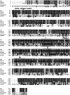
Figure 1
Figure 1
Amino-acid sequence alignment of PRMT3 from rat, mouse, human, fly, and fission yeast. Identical amino acids are shown in black outline and similar amino acids are shown in gray outline. Conserved protein methyltransferase motifs I, post-I, II, and III are boxed in gray. The putative C2H2 zinc-finger motif of PRMT3 is boxed in white. An asterisk is present under the canonical cysteine and histidine residues of the zinc-finger motif. Alignments and shading were generated using Clustalw and Boxshade, both available via the World Wide Web. The GenBank accession numbers used for the alignment are as follows: NP_446009 for rat; NP_598501 for mouse; XP_058460 for human; AAO24922 for Drosophila; CAA17825 for S. pombe.
Figure 2
Figure 2
Cytoplasmic localization of fission yeast PRMT3. (A) Whole-cell extracts were prepared from S. pombe strains expressing PRMT1-GFP and PRMT3-GFP, and immunoblotted with an affinity-purified GFP antibody. (B) Cells expressing carboxy-terminal-tagged PRMT1 and PRMT3 were grown in YES medium. Live S. pombe cells were analyzed by Nomarski microscopy (a, c) and by fluorescence microscopy (b, d) for PRMT3-GFP (a, b) and PRMT1-GFP (c, d) localization.
Figure 3
Figure 3
TAP of PRMT3 from fission yeast. Proteins copurified with PRMT3 by TAP (lane 2) were resolved using a Bis-Tris 4–12% gradient SDS–PAGE and analyzed by silver staining. The result for an identically treated extract from wild-type untagged S. pombe is shown in lane 1. Molecular weight markers are shown on the right in kilodaltons (kDa). The positions of PRMT3 and the 40S ribosomal protein S2 (rpS2) identified by mass spectrometry are indicated on the left.
Figure 4
Figure 4
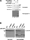
The 40S ribosomal protein S2 (rpS2) is asymmetrically methylated by PRMT3. (A) A 1 μg portion of recombinant arginine–glycine-rich substrate (RGG)n (lanes 1 and 2) and rpS2 (lanes 3 and 4) were incubated with (lanes 2 and 4) or without (lanes 1 and 3) recombinant PRMT3 (1 μg) in the presence of 1 μCi of [3H]AdoMet for 3 h at 32°C. Proteins were separated by SDS–PAGE and stained with Coomassie blue (upper panel). The stained gel was dried and exposed for fluorography for 72 h (lower panel). The positions of recombinant GST-PRMT3, GST-rpS2, and GST-(RGG)n are shown on the left of the upper panel. (B) Total cell extracts were prepared from log-phase yeast cultures inoculated from two independent colonies. Immunoblots of total protein lysates prepared from wild-type (lanes 1, 2, 7, 8, 9, and 14), prmt1-disrupted (ΔPRMT1; lanes 3, 4, 10, and 11), and prmt3-disrupted (ΔPRMT3; lanes 5, 6, 12, and 13) yeast strains that either expressed a C-terminal GFP-tagged rpS2 (lanes 1–6 and 8–13) or untagged rpS2 (lanes 7 and 14). An affinity-purified GFP antibody (anti-GFP) was used to detect the total rpS2-GFP protein (left panel) and an affinity-purified antibody specific for asymmetrically dimethylated arginines (anti-ASYM24) was used to detect the methylated form of rpS2-GFP. The position of rpS2-GFP is shown on the right whereas the molecular weight markers are shown on the left in kilodaltons (kDa).
Figure 5
Figure 5
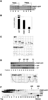
A fraction of fission yeast PRMT3 associates with ribosomes and cosediments with free 40S subunits. (A) S. pombe cells expressing a C-terminal GFP-tagged PRMT3 were treated with cycloheximide, lysed, and centrifuged through a 20% (w/v) sucrose cushion. In all, 0.125, 0.25, 0.5, and 1% of the total cell lysate (Input; lanes 1–4) and 2.5, 5, 10, and 20% of the ribosome pellet (Pellet; lanes 5–8) were subjected to SDS–PAGE and immunoblot analysis using anti-GFP, anti-actin, and anti-60S ribosomal protein L7 (rpL7). The 60S rpL7 protein could be detected in the input upon longer exposures (data not shown). (B) Quantitation of ribosome-associated rpL7, PRMT3, and actin. Values are expressed in a graphical form as percentage of input. Data were quantified for at least three independent experiments (see Materials and methods). (C) Polysome profiles of ribosomes isolated from wild-type (WT) and PRMT3-GFP-expressing cells resolved using a 5–45% sucrose gradient. The positions of free small (40S) and large (60S) ribosomal subunits, monosomes (80S), and polysomes (2–6) are indicated in the WT profile (upper panel). (D) Fractions from the gradient isolated from the PRMT3-GFP-expressing strain shown in (A) were collected, the proteins were precipitated using trichloroacetic acid, and analyzed by immunoblotting. The positions of the 40S, 60S, 80S monosomes, and P2 polysomes are indicated at the top. (E) Extracts from PRMT3-expressing cells were separated by sucrose gradient as in (C) but centrifuged for 12 h. The positions of free small (40S) and large (60S) ribosomal subunits are indicated. (F) Fractions from the gradient resolved in (E) were precipitated and the proteins were analyzed by immunoblotting.
Figure 6
Figure 6
Human PRMT3 and the 70-kDa S6 kinase 1 show similar distributions along ribosome profiles. (A) Polysome profiles of ribosomes isolated from HeLa cells were separated using a 5–45% sucrose gradient. The positions of free small (40S) and large (60S) ribosomal subunits, monosomes (80S), and polysomes (2–6) are indicated. (B) Fractions from the gradient shown in (A) were collected and the proteins were precipitated using trichloroacetic acid. Proteins were analyzed by immunoblotting using antibodies to detect human PRMT3 (hPRMT3), the ribosomal protein S6 kinase 1 (S6K1), phospho-Thr389-S6K1 (P-S6K1), human 40S ribosomal protein S6 (hrpS6), and the human 60S ribosomal protein L7 (hrpL7). The positions of the 40S, 60S, 80S monosomes, and polysomes are indicated at the top.
Figure 7
Figure 7
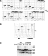
Imbalance in the free 40S:60S ratio in prmt3-disrupted cells. (A) Polysome profiles of ribosomes isolated from wild-type (WT; upper left panel), prmt1-disrupted (ΔPRMT1; upper right panel), and prmt3-disrupted (ΔPRMT3; bottom panels) cells were separated using 5–45% sucrose gradients. The positions of free small (40S) and large (60S) ribosomal subunits and monosomes (80S) are indicated. (B) Total ribosomes were isolated from wild-type (WT) and prmt3-disrupted (ΔPRMT3) cells and dissociated into 40S and 60S subunits by the addition of 40 mM EDTA. The dissociated ribosomal subunits were resolved onto 5–45% sucrose gradients. (C) Normal pre-rRNA processing in prmt3-disrupted cells. Exponentially growing cultures of wild-type and prmt3-disrupted S. pombe were pulse-labeled with 200 μCi of [methyl-3H]methionine for 3 min, and then chased for 1.5, 3, and 6 min after the addition of unlabeled methionine. Total cellular RNA was extracted from cells at each time point and 10 000 cpm was analyzed by formaldehyde–agarose gel. The positions of the different pre-rRNAs and mature rRNAs are indicated on the right.

References

    1. Abramovich C, Yakobson B, Chebath J, Revel M (1997) A protein-arginine methyltransferase binds to the intracytoplasmic domain of the IFNAR1 chain in the type I interferon receptor. EMBO J 16: 260–266 - PMC - PubMed
    1. Akiyoshi Y, Clayton J, Phan L, Yamamoto M, Hinnebusch AG, Watanabe Y, Asano K (2001) Fission yeast homolog of murine Int-6 protein, encoded by mouse mammary tumor virus integration site, is associated with the conserved core subunits of eukaryotic translation initiation factor 3. J Biol Chem 276: 10056–10062 - PubMed
    1. All-Robyn JA, Brown N, Otaka E, Liebman SW (1990) Sequence and functional similarity between a yeast ribosomal protein and the Escherichia coli S5 ram protein. Mol Cell Biol 10: 6544–6553 - PMC - PubMed
    1. Bedford MT, Frankel A, Yaffe MB, Clarke S, Leder P, Richard S (2000) Arginine methylation inhibits the binding of proline-rich ligands to Src homology 3, but not WW, domains. J Biol Chem 275: 16030–16036 - PubMed
    1. Boisvert FM, Cote J, Boulanger MC, Cleroux P, Bachand F, Autexier C, Richard S (2002) Symmetrical dimethylarginine methylation is required for the localization of SMN in Cajal bodies and pre-mRNA splicing. J Cell Biol 159: 957–969 - PMC - PubMed

Publication types

MeSH terms