

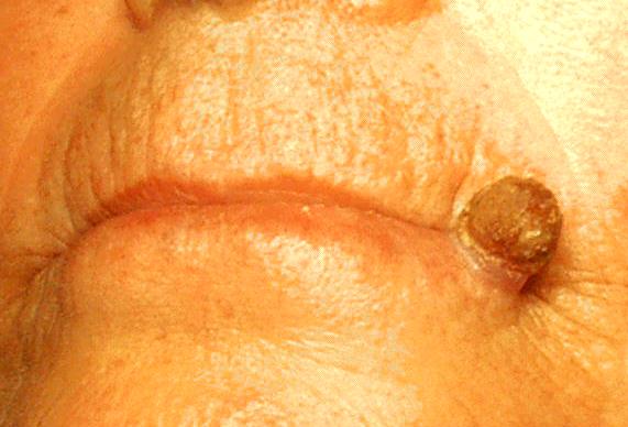
Cutaneous horns: are these lesions as innocent as they seem to be?
- PMID: 15176977
- PMCID: PMC421749
- DOI: 10.1186/1477-7819-2-18
Cutaneous horns: are these lesions as innocent as they seem to be?
Abstract
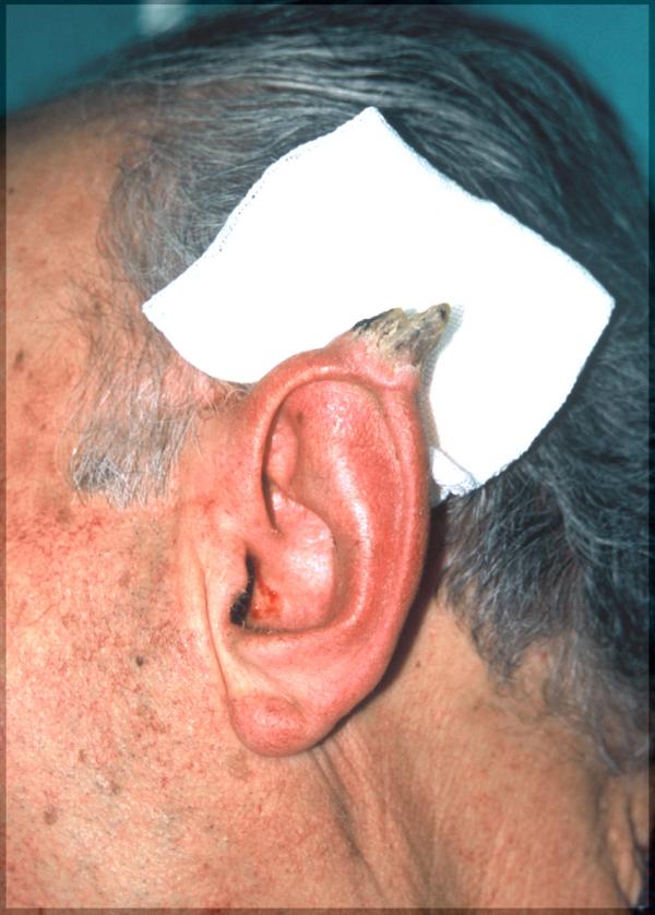
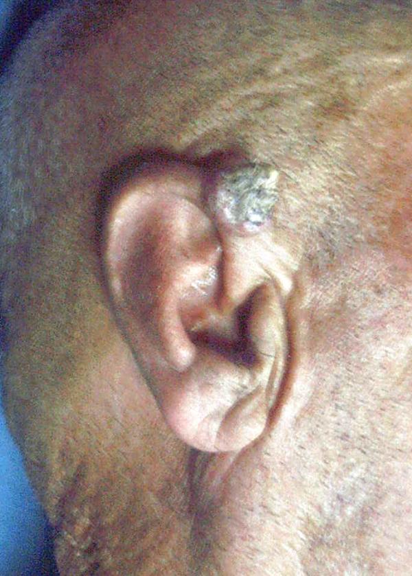
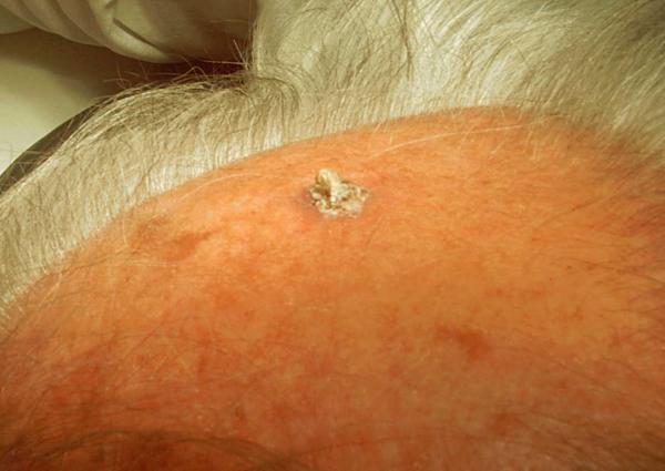
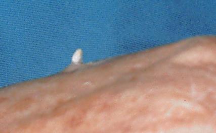
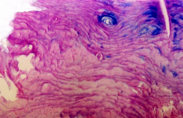
Background: Cutaneous horns (cornu cutaneum) are uncommon lesions consisting of keratotic material resembling that of an animal horn. Cutaneous horn may arise from a wide range of the epidermal lesions, which may be benign, premalignant or malignant.
Patients and methods: In this respective study, we describe our experience of eleven patients with cutaneous horn treated at our centre between January 2000 and January 2004. The clinical, pathological and treatment details were extracted from the case records. Data is presented as frequency distribution.
Results: There were 8 male and 3 female patients with a median age of 57 years. Most of the lesions were located on the ear, hand and scalp. Surgical resection was carried out in all the lesions. There were two cases of squamous cell carcinoma, and one case of basal cell carcinoma, other 8 cases were benign. None of the lesions recurred and no adjuvant treatment was given to any of the malignant lesions.
Conclusion: Cutaneous horn is a clinical diagnosis that refers to a conical projection above the surface of the skin. The lesions typically occurs in sun exposed areas, particularly the face, ear, nose, forearms, and dorsum of hands. Even though our 60% of the cutaneous horns are benign possibility of skin cancer should always be kept in mind.
Figures
References
-
- Korkut T, Tan NB, Oztan Y. Giant cutaneous horn: a patient report. Ann Plast Surg. 1997;39:654–655. - PubMed
-
- Yu RC, Pryce DW, Macfarlane AW, Stewart TW. A histopathological study of 643 cutaneous horns. Br J Dermatol. 1991;124:449–452. - PubMed
-
- Michal M, Bisceglia M, Di Mattia A, Requena L, Fanburg-Smith JC, Mukensnabl P, Hes O, Cada F. Gigantic cutaneous horns of the scalp: lesions with a gross similarity to the horns of animals: a report of four cases. Am J Surg Pathol. 2002;26:789–794. doi: 10.1097/00000478-200206000-00014. - DOI - PubMed
LinkOut - more resources
Full Text Sources
