. 2004 Jun 3:5:3.
doi: 10.1186/1471-2199-5-3.

The expression of HSP83 genes in Leishmania infantum is affected by temperature and by stage-differentiation and is regulated at the levels of mRNA stability and translation

Affiliations

The expression of HSP83 genes in Leishmania infantum is affected by temperature and by stage-differentiation and is regulated at the levels of mRNA stability and translation

Ruth Larreta et al. BMC Mol Biol. .

Abstract

Background: Exposure of Leishmania promastigotes to the temperature of their mammalian hosts results in the induction of a typical heat shock response. It has been suggested that heat shock proteins play an important role in parasite survival and differentiation.

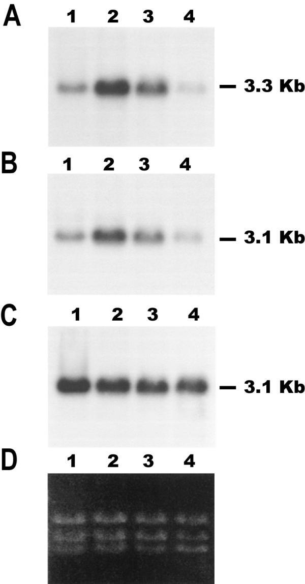
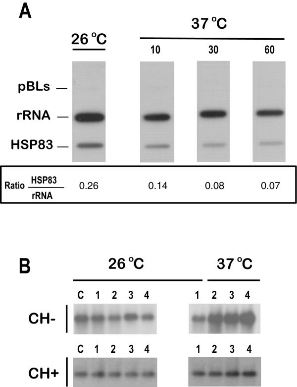
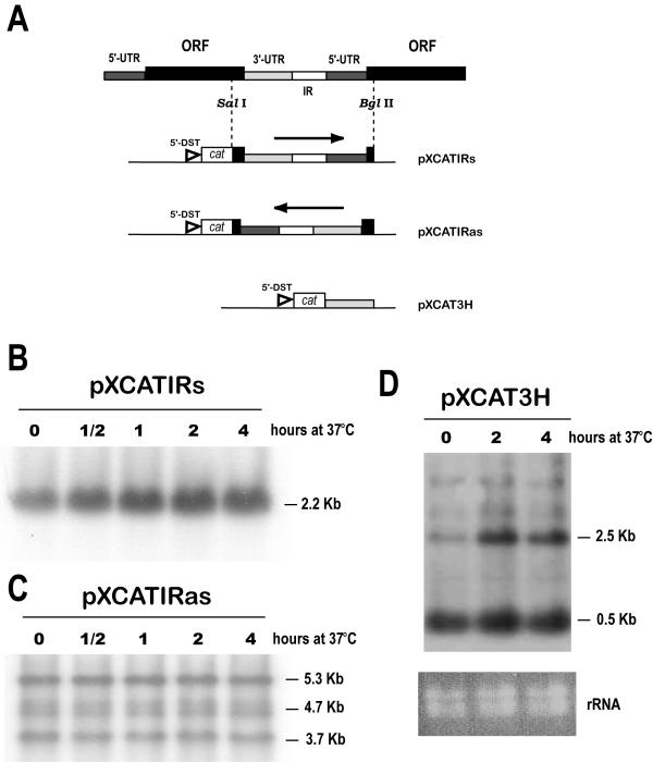
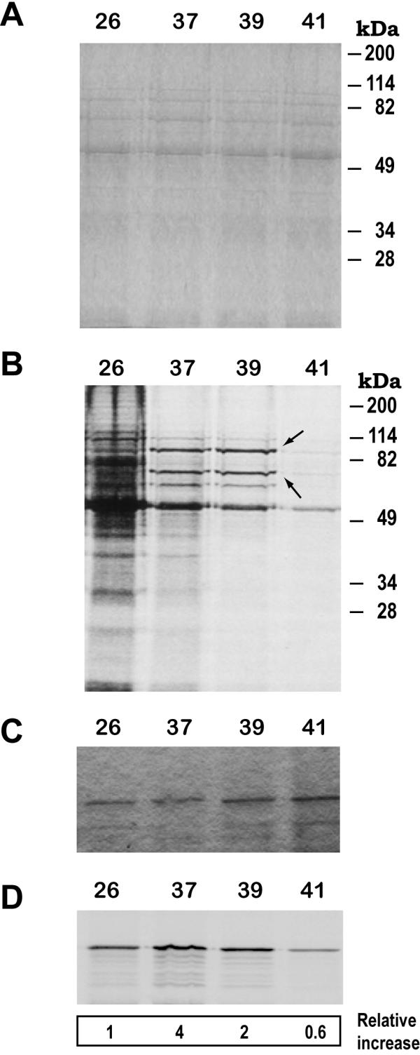
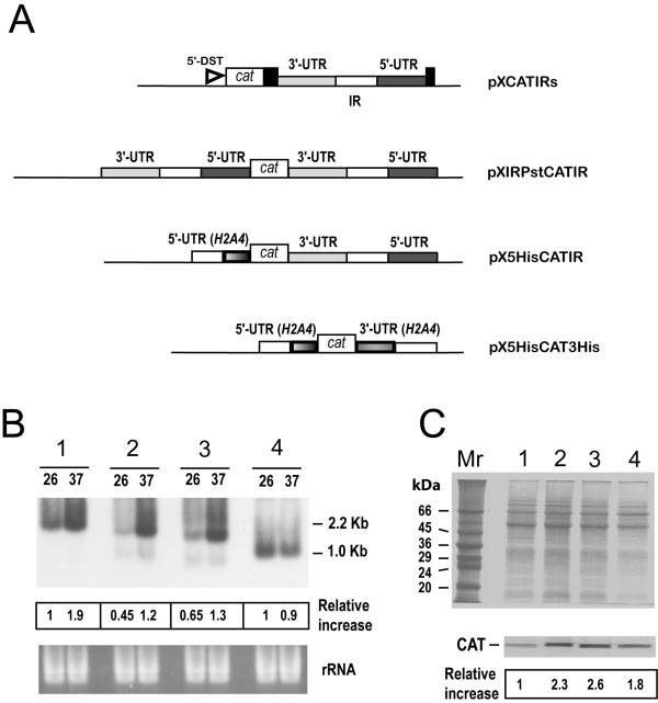
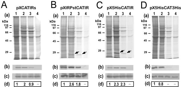
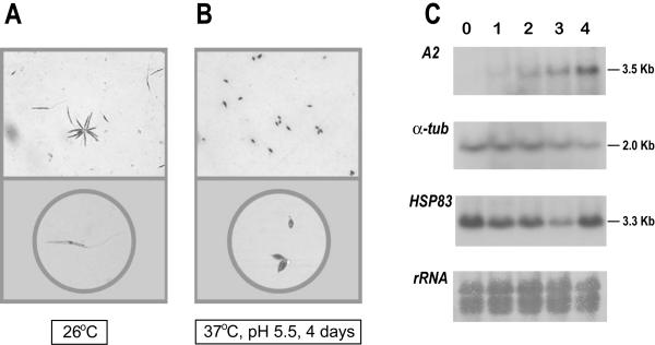
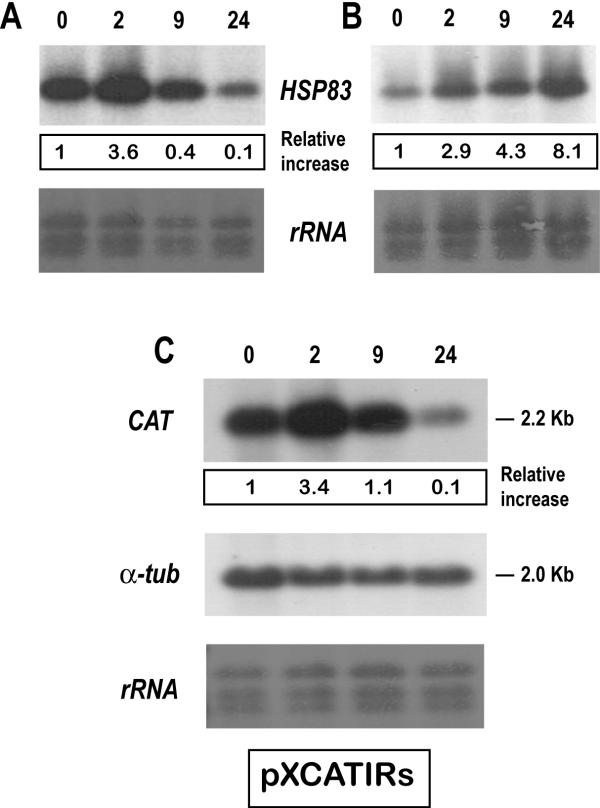
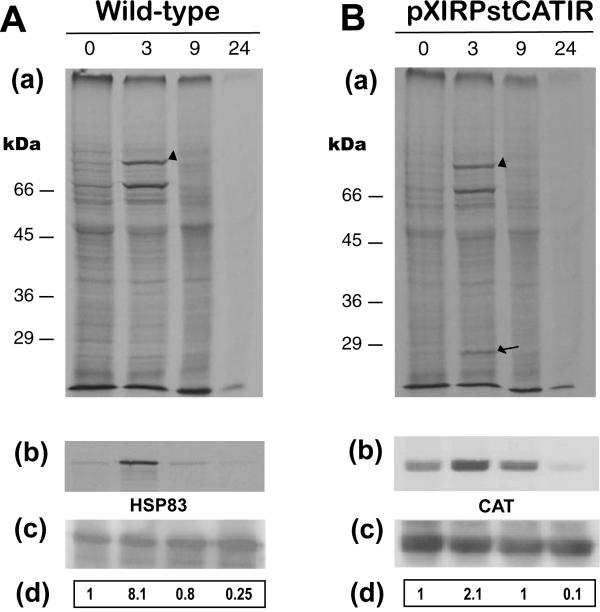
Results: Here we report the studies on the expression of the heat shock protein 83 (HSP83) genes of Leishmania infantum. Confirming previous observations for other Leishmania species, we found that the L. infantum HSP83 transcripts also show a temperature-dependent accumulation that is controlled by a post-transcriptional mechanism involving sequences located in the 3'-untranslated region (3'-UTR). However, contrary to that described for L. amazonensis, the accumulation of the HSP83 transcripts in L. infantum is dependent on active protein synthesis. The translation of HSP83 transcripts is enhanced during heat shock and, as first described in L. amazonensis, we show that the 3'-UTR of the L. infantum HSP83 gene is essential for this translational control. Measurement of the steady-state levels of HSP83 transcripts along the promastigote-to-amastigote differentiation evidenced a specific profile of HSP83 RNAs: after an initial accumulation of HSP83 transcripts observed short after (2 h) incubation in the differentiation conditions, the amount of HSP83 RNA decreased to a steady-state level lower than in undifferentiated promastigotes. We show that this transient accumulation is linked to the presence of the 3'-UTR and flanking regions. Again, an 8-fold increase in translation of the HSP83 transcripts is observed short after the initiation of the axenic differentiation, but it is not sustained after 9 h.

Conclusions: This transient expression of HSP83 genes could be relevant for the differentiation of Leishmania, and the underlying regulatory mechanism may be part of the developmental program of this parasite.


Figures

Figure 1
Figure 1
Temperature-dependent accumulation of transcripts for HSP83 and HSP70. RNA samples were prepared from promastigotes grown at 26°C (lanes 1) or incubated for 2 h at 37°C (lanes 2), 39°C (lanes 3), or 41°C (lanes 4), and analyzed by Northern blotting. Northern blots were hybridized with the L. infantum HSP83 3'-UTR (panel A). The same blot was stripped and rehybridized with probes for the L. infantum HSP70 3'-UTR-I (panel B) and the L. infantum HSP70 3'-UTR-II (panel C) [9]. Panel D shows an ethidium bromide staining of the RNA samples loaded on agarose gel prior to transfer.
Figure 2
Figure 2
The regulation of the HSP83 gene expression occurs at the post-transcriptional level. (A) Promastigotes of L. infantum were grown at 26°C, and parallel cultures were incubated at 37°C during 10, 30, and 60 min. Run-on transcripts, labeled with [32P]UTP, were hybridized to slot blots containing 5 μg of linearized plasmid DNAs. The following plasmids were used: pBLs, pBluescript; rRNA, pRIB (plasmid containing a 590-bp fragment of the L. infantum 24Sα rDNA gene [9]); HSP83, pE2 (plasmid containing a fragment of the 3'-UTR of L. infantum HSP83 gene). The values of the hybridization intensity were measured by densitometric analysis of the autoradiographs, and the ratio HSP83/rRNA is shown. Data represent the average of two separate experiments. (B) RNA samples were prepared from promastigotes cultures grown at either 26°C or 37°C for 0 h (lanes 1), 1 h (lanes 2), 2 h (lanes 3) and 4 h (lanes 4) in the presence (CH+) or absence (CH-) of cycloheximide. The drug was added 1 h prior to start the temperature treatment. Lanes C contain RNA samples prepared from parasite cultures before starting the drug treatment. The Northern blots were hybridized with a specific probe for the L. infantum HSP83 3'-UTR.
Figure 3
Figure 3
Role of the 3'-UTR of the HSP83 gene in the temperature-dependent accumulation of transcripts. (A) Schematic representation of a portion of the L. infantum HSP83 gene cluster, showing the two most 5' genes of the cluster. The SalI+BglII genomic fragment was cloned in both orientations into pXcat plasmid to yield constructs pXCATIRs (sense) and pXCATIRas (antisense). Construct pXCAT3H was obtained by insertion of the 3'-UTR of HSP83 gene downstream of the cat ORF (see Materials and methods for cloning details). The position of the L. major DST sequence (5'-DST) [25], providing the miniexon addition site for the cat transcripts, is shown. Both pXCATIRs-transfected (panel B) and pXCATIRas-transfected (panel C) promastigotes were incubated either at 26°C or 37°C for 30 min, 1 h, 2 h, and 4 h; afterwards, RNA was extracted and used to prepare Northern blots that were hybridized with a cat probe. (D) RNA was extracted from pXCAT3H-transfected promastigotes after incubation at 37°C for various time points (0, 2, and 4 h). The Northern blot was hybridized with the cat probe. At the bottom, panel rRNA shows an ethidium bromide staining of the RNA samples loaded on the agarose gel.
Figure 4
Figure 4
The translation of HSP83 is enhanced in L. infantum promastigotes by incubation at 37°C and 39°C. L. infantum promastigotes were grown at either 26°C, 37°C, 39°C, or 41°C for 2 h in the presence of [35S]methionine/cysteine. Protein samples corresponding to 106 cells were separated over 10% SDS-PAGE and the gel was stained with Coomassie blue (panel A) and, subsequently, the labeled protein bands were revealed by autoradiography (panel B). On panel B, the position of putative HSP83 and HSP70 bands is indicated by arrows. In parallel, lysates from [35S]-labeled promastigotes incubated at the different temperatures were used to immunoprecipitate the HSP83 with specific antibodies. Immunoprecipitates were subjected to SDS-PAGE (panel C). After drying, the autoradiography of the gel was obtained (panel D). The ratio (expressed as relative increase) between incorporation of [35S] and the intensity of the protein band (stained by Coomassie blue) was determined. The data are averages of three independent experiments.
Figure 5
Figure 5
Analysis of the expression of cat transcripts and CAT protein in the different transfected Leishmania cell lines. (A) Schematic maps of the constructs. The CAT coding gene (open box) was cloned between different 5'-UTRs and 3'-UTRs. Where not indicated, the UTRs are derived from L. infantum HSP83 genes. Along with the UTRs, the corresponding IRs (open rectangles) were also included. The position of the L. major DST sequence (5'-DST) in construct pXCATIRs is indicated. (B) RNA samples from promastigotes transfected with pXCATIRs (1), pXIRPstCATIR (2), pX5HisCATIR (3), or pX5HisCAT3His (4), incubated for 2 h either at 26°C (lanes 26) or at 37°C (lanes 37), were assayed by Northern analysis using a cat probe. The bottom panel shows an ethidium bromide staining of the RNA samples loaded on the gel prior to transfer. The intensities of the hybridization bands were evaluated by densitometric analysis and expressed as a relative increase (giving an arbitrary value of 1 to the intensity found in lane 26 of pXCATIRs-sample). (C) SDS-PAGE and Coomassie blue staining of protein extracts (4 × 10⁶ cells per lane) from different promastigote lines grown at 26°C. The stable transfections used are: pXCATIRs (lane 1), pXIRPstCATIR (lane 2), pX5HisCATIR (lane 3), and pX5HisCAT3His (lane 4). The bottom panel shows a Western blot analysis; proteins were transferred to a nitrocellulose membrane and incubated with an anti-CAT antibody (dilution 1:1000). The intensity of the bands was quantified by densitometric scanning and the normalized relative signals (relative increase) are shown. Data represent mean values from two independent experiments.
Figure 6
Figure 6
The HSP83 3'-UTR is essential for enhanced translation during heat shock. L. infantum promastigotes transfected with constructs pXCATIRs (A), pXIRPstCATIR (B), pX5HisCATIR (C), or pX5HisCAT3His (D) were metabolically labeled for 2 h at 26°C (lanes 1), 37°C (lanes 2), 39°C (lanes 3), or 41°C (lanes 4) in the presence of [35S]methionine/cysteine. Proteins (4 × 10⁶ cells per lane) were separated by SDS-PAGE and the corresponding autoradiographies are shown (panels a). In parallel, cells were harvested and lysed, and the CAT protein was immunoprecipited using a specific antibody. Panel b shows the immunoprecipitation followed by autoradiography. Panel c shows the immunoprecipitation followed by Western blot using an anti-CAT antibody. The label intensity of the immunoprecipitated CAT protein (panels b) was measured by densitometric scanning and normalized relative to the total amount of immunoprecipitated CAT protein as determined by Western blot (panels c). The relative signals are shown in panels d. Data represent mean values derived from at least two independent experiments.
Figure 7
Figure 7
Differentiation of promastigotes to amastigotes in axenic conditions. Morphology of L. infantum promastigotes (A) and amastigote-like forms (B). Logarithmic promastigotes and amastigote-like forms (after 4 days of differentiation) were microscopically evaluated. Parasites were placed on slides and, after air dried, stained with DADE® Diff-Quik® (Baxter Diagnostics, Dudingen, Switzerland). The magnifications used to take the micrographs were: 400 × (top panels) and 630 × (bottom panels). (C) Expression of A2, α-tubulin, and HSP83 transcripts during differentiation of promastigotes to amastigotes. Promastigotes (lane 0) were differentiated to amastigotes (see Materials and methods for details). Aliquots of 5 × 10⁷ cells were taken every 24 h (day 1, 2, 3, and 4) and used to extract RNA. The Northern blot was probed with the L. donovani A2 gene [26] and, after stripping, the blot was reprobed with the T. cruzi α-tubulin gene [38], and the 3'-UTR of the L. infantum HSP83 gene. A methylene blue staining of the blot is shown at the bottom (panel rRNA).
Figure 8
Figure 8
Steady-state levels of HSP83 transcripts fluctuate during promastigote-to-amastigote differentiation. L. infantum promastigotes were cultured under amastigote growth conditions (panel A) or incubated at 37°C (panel B). RNA samples were prepared at various time points within the treatments (0, 2, 9 and 24 h), transferred onto a nylon membrane and hybridized with the HSP83 3'-UTR probe. C, pXCATIRs-transfected promastigotes were subjected to amastigote growth conditions for 0, 2, 9 and 24 h. The Northern blot was hybridized with the cat probe, and subsequently with the T. cruzi α-tubulin. Bottom panels show methylene blue staining of blots. The autoradiographs were quantified by densitometric scanning and normalized to the intensity at 26°C (arbitrary value 1). The data (relative increase) correspond to mean values derived from at least two independent experiments.
Figure 9
Figure 9
The translation rate of the L. infantum HSP83 fluctuates during promastigote-to-amastigote differentiation. Wild-type (A) or pXIRPstCATIR-transfected (B) promastigotes were incubated during 0, 3, 9 and 24 h in amastigote growth conditions. The parasites were metabolically labeled with [35S]methionine/cysteine for the last 2 h of treatment. Proteins were extracted and analyzed by 10% SDS-PAGE, followed by autoradiography of the dried gels (panels a). The HSP83 band is marked with an arrowhead and the CAT band is marked with an arrow. The numbers at the left indicate the mobility and relative molecular weights (in kDa) of protein molecular weight standards. In parallel, cells were lysed and used to immunoprecipitate either the HSP83 (A) or CAT (B) using specific antibodies. Panels b show the immunoprecipitations followed by autoradiography; panels c show the immunoprecipitations followed by Western blotting. The label intensities of the immunoprecipitated proteins (panels b) were measured by densitometric scanning and normalized relative to the total amount of immunoprecipitated proteins (panels c). The relative signals are shown in panels d. Data represent mean values derived from at least three independent experiments.

Similar articles

Cited by

References

    1. Zilberstein D, Shapira M. The role of pH and temperature in the development of Leishmania parasites. Annu Rev Microbiol. 1994;48:449–470. doi: 10.1146/annurev.mi.48.100194.002313. - DOI - PubMed
    1. Saar Y, Ransford A, Waldman E, Mazareb S, Amin-Spector S, Plumblee J, Turco SJ, Zilberstein D. Characterization of developmentally-regulated activities in axenic amastigotes of Leishmania donovani. Mol Biochem Parasitol. 1998;95:9–20. doi: 10.1016/S0166-6851(98)00062-0. - DOI - PubMed
    1. Somanna A, Mundodi V, Gedamu L. In vitro cultivation and characterization of Leishmania chagasi amastigote-like forms. Acta Trop. 2002;83:37–42. doi: 10.1016/S0001-706X(02)00054-2. - DOI - PubMed
    1. Lawrence F, Robert-Gero M. Induction of heat shock and stress proteins in promastigotes of three Leishmania species. Proc Natl Acad Sci USA. 1985;82:4414–4417. - PMC - PubMed
    1. Stiles JK, Hicock PI, Shah PH, Meade JC. Genomic organization, transcription, splicing and gene regulation in Leishmania. Ann Trop Med Parasitol. 1999;93:781–807. doi: 10.1080/00034989957781. - DOI - PubMed

Publication types

MeSH terms

LinkOut - more resources