

Rapid and sequential movement of individual chromosomal loci to specific subcellular locations during bacterial DNA replication
- PMID: 15178755
- PMCID: PMC438963
- DOI: 10.1073/pnas.0402606101
Rapid and sequential movement of individual chromosomal loci to specific subcellular locations during bacterial DNA replication
Abstract
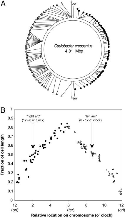
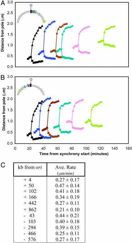
The chromosomal origin and terminus of replication are precisely localized in bacterial cells. We examined the cellular position of 112 individual loci that are dispersed over the circular Caulobacter crescentus chromosome and found that in living cells each locus has a specific subcellular address and that these loci are arrayed in linear order along the long axis of the cell. Time-lapse microscopy of the location of the chromosomal origin and 10 selected loci in the origin-proximal half of the chromosome showed that during DNA replication, as the replisome sequentially copies each locus, the newly replicated DNA segments are moved in chronological order to their final subcellular destination in the nascent half of the predivisional cell. Thus, the remarkable organization of the chromosome is being established while DNA replication is still in progress. The fact that the movement of these 10 loci is, like that of the origin, directed and rapid, and occurs at a similar rate, suggests that the same molecular machinery serves to partition and place many, if not most, chromosomal loci at defined subcellular sites.
Figures


Comment in
-
Linear ordering and dynamic segregation of the bacterial chromosome.Proc Natl Acad Sci U S A. 2004 Jun 22;101(25):9175-6. doi: 10.1073/pnas.0403722101. Epub 2004 Jun 15. Proc Natl Acad Sci U S A. 2004. PMID: 15199189 Free PMC article. Review. No abstract available.
Similar articles
-
Coordination between chromosome replication, segregation, and cell division in Caulobacter crescentus.J Bacteriol. 2006 Mar;188(6):2244-53. doi: 10.1128/JB.188.6.2244-2253.2006. J Bacteriol. 2006. PMID: 16513754 Free PMC article.
-
Caulobacter requires a dedicated mechanism to initiate chromosome segregation.Proc Natl Acad Sci U S A. 2008 Oct 7;105(40):15435-40. doi: 10.1073/pnas.0807448105. Epub 2008 Sep 29. Proc Natl Acad Sci U S A. 2008. PMID: 18824683 Free PMC article.
-
Compaction and transport properties of newly replicated Caulobacter crescentus DNA.Mol Microbiol. 2011 Dec;82(6):1349-58. doi: 10.1111/j.1365-2958.2011.07899.x. Epub 2011 Nov 16. Mol Microbiol. 2011. PMID: 22085253
-
Escherichia coli and its chromosome.Trends Microbiol. 2008 May;16(5):238-45. doi: 10.1016/j.tim.2008.02.003. Epub 2008 Apr 9. Trends Microbiol. 2008. PMID: 18406139 Review.
-
Cell cycle regulation in Caulobacter: location, location, location.J Cell Sci. 2007 Oct 15;120(Pt 20):3501-7. doi: 10.1242/jcs.005967. J Cell Sci. 2007. PMID: 17928306 Review.
Cited by
-
Organization and segregation of bacterial chromosomes.Nat Rev Genet. 2013 Mar;14(3):191-203. doi: 10.1038/nrg3375. Epub 2013 Feb 12. Nat Rev Genet. 2013. PMID: 23400100 Free PMC article. Review.
-
Characterization of chromosomal and megaplasmid partitioning loci in Thermus thermophilus HB27.BMC Genomics. 2015 Apr 18;16(1):317. doi: 10.1186/s12864-015-1523-3. BMC Genomics. 2015. PMID: 25909452 Free PMC article.
-
HdaB: a novel and conserved DnaA-related protein that targets the RIDA process to stimulate replication initiation.Nucleic Acids Res. 2020 Mar 18;48(5):2412-2423. doi: 10.1093/nar/gkz1193. Nucleic Acids Res. 2020. PMID: 31875223 Free PMC article.
-
Why and how bacteria localize proteins.Science. 2009 Nov 27;326(5957):1225-8. doi: 10.1126/science.1175685. Science. 2009. PMID: 19965466 Free PMC article. Review.
-
The three-dimensional architecture of a bacterial genome and its alteration by genetic perturbation.Mol Cell. 2011 Oct 21;44(2):252-64. doi: 10.1016/j.molcel.2011.09.010. Mol Cell. 2011. PMID: 22017872 Free PMC article.
References
Publication types
MeSH terms
Substances
Grants and funding
LinkOut - more resources
Full Text Sources
Other Literature Sources
