Comparative Study
. 2004 Jun 15;101(24):8900-5.
doi: 10.1073/pnas.0401904101. Epub 2004 Jun 3.

Visualization of ribosome-recycling factor on the Escherichia coli 70S ribosome: functional implications

Affiliations
Comparative Study

Visualization of ribosome-recycling factor on the Escherichia coli 70S ribosome: functional implications

Rajendra K Agrawal et al. Proc Natl Acad Sci U S A. .

Abstract

After the termination step of protein synthesis, a deacylated tRNA and mRNA remain associated with the ribosome. The ribosome-recycling factor (RRF), together with elongation factor G (EF-G), disassembles this posttermination complex into mRNA, tRNA, and the ribosome. We have obtained a three-dimensional cryo-electron microscopic map of a complex of the Escherichia coli 70S ribosome and RRF. We find that RRF interacts mainly with the segments of the large ribosomal subunit's (50S) rRNA helices that are involved in the formation of two central intersubunit bridges, B2a and B3. The binding of RRF induces considerable conformational changes in some of the functional domains of the ribosome. As compared to its binding position derived previously by hydroxyl radical probing study, we find that RRF binds further inside the intersubunit space of the ribosome such that the tip of its domain I is shifted (by approximately 13 A) toward protein L5 within the central protuberance of the 50S subunit, and domain II is oriented more toward the small ribosomal subunit (30S). Overlapping binding sites of RRF, EF-G, and the P-site tRNA suggest that the binding of EF-G would trigger the removal of deacylated tRNA from the P site by moving RRF toward the ribosomal E site, and subsequent removal of mRNA may be induced by a shift in the position of 16S rRNA helix 44, which harbors part of the mRNA.


Figures

Fig. 1.
Fig. 1.
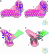
Stereoview representations of the three-dimensional cryo-EM structure of the 70S ribosome–RRF complex. (a) 70S ribosome, showing the shoulder side of the 30S subunit (yellow) on the left, and L7/L12-stalk side of the 50S subunit (blue) on the right to reveal the binding position of RRF (red). (b) Computationally separated RRF density superposed onto the 30S subunit. (c) RRF density superposed onto the 50S subunit. hd, head; sh, shoulder; sp, spur; pt, platform; L1, L1-protein protuberance; CP, central protuberance; St, L7/L12 stalk; Sb, stalk-base; SRL, α-sarcin-ricin loop; h and H followed by numbers identify the 16S and 23S rRNA helices, respectively; I and II, domains of RRF; asterisk, density of a tRNA in the P/E state.
Fig. 2.
Fig. 2.
Structural changes of the ribosome upon RRF binding. (a) Maps of RRF-bound ribosome (semitransparent blue) and the naked (control) ribosome (purple) have been superimposed. The 50S subunits are shown from the intersubunit-face side, and portions corresponding to the L7/L12 stalk-base region (Sb) and the Stalk (St) are magnified in Inset. Densities corresponding to RRF (red) and RRF-binding-associated conformational change (green, see text), which is visible only at lower threshold values, are also shown. (b) Same maps as in a, but transparencies have been flipped to reveal the conformational change associated with the 23S rRNA helix 69 (H69). Superimposed maps have been rotated with respect to their orientation in a, around a horizontal axis, by ≈35°, such that CP of the 50S subunit moves closer to the viewer. The helix 69 portion has been magnified in Inset. (c) Superimposed 30S subunits of the RRF-bound ribosome (semitransparent yellow) and naked ribosome (orange), shown from the intersubunit-face side. The helix 44 (h44) region of the 16S rRNA is magnified in Inset. In b and c Insets, ribosome masses from the far plane have been computationally removed for visual clarity. Arrows indicate the direction of movements of ribosomal mass upon RRF binding. Asterisk in a indicates a significant conformational change in the L7/L12 stalk, which becomes a more defined, thick, and extended structure upon RRF binding, as also seen previously in the case of EF-G (37). Locations of two bridges (B2a and B3) before and after RRF binding are indicated as ovals with solid and broken lines, respectively. I and II, two structural domains of RRF. All other landmarks are the same as in Fig. 1.
Fig. 3.
Fig. 3.
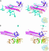
Fitting of atomic structure of RRF into the cryo-EM density. (a) Stereo-view representation of the fitting of the x-ray structure of T. maritima RRF (domain I, pink; and domain II, violet) into the cryo-EM density (semitransparent red). In most part, the x-ray structure of RRF is embedded within the cryo-EM density, recognizable by altered hues of color for the two RRF domains (reddish pink for domain I, magenta for domain II), whereas very small portions of the x-ray structure that stick out of the cryo-EM density are visible in their original (pink or violet) colors. The cryo-EM density of RRF is shown at a lower threshold value as compared to the value used in Figs. 1 and 2. (b) Same as a, but shown from the intersubunit space side, with L7/L12 stalk facing the viewer. (c) Alternative fitting of RRF atomic structure (gray) into the cryo-EM map shown from the same view as in b. In this fitting, domain II of RRF is oriented into a weaker mass of density (semitransparent green, see text), toward the L7/L12 stalk-base region. This fitting yields a significantly lower cross-correlation value (0.51 vs. 0.77 for the fitting shown in a and b). For the purpose of comparison, only Cα backbones of RRF x-ray structure are shown in b and c. The orientations of the 70S ribosome (semitransparent surfaces), with RRF density (red), are shown as thumbnails on the lower right. All of the landmarks are the same as in Fig. 2.
Fig. 4.
Fig. 4.
Sites of interaction between RRF and ribosome. X-ray crystallographic structures of both ribosomal subunits (28, 41) were fitted into the cryo-EM map of the RRF-ribosome complex. (a) The 23S rRNA helices (light blue) of the 50S ribosome that directly interact with (or lie within 3 Å of) RRF are shown. Domains I (magenta) and II (purple) of the RRF atomic structure are assigned the same colors as in Fig. 3 a and b, with conserved (red) and semiconserved (orange) residues highlighted. The RRF residues making direct contact with rRNA are shown with side chains (red). Ribosomal RNA residues making direct contacts with RRF are highlighted as beads in corresponding color. (b) Regions of ribosome that lie within 5–10 Å distant [except for helix 43 (H43) of the 23S rRNA, see below], and the position of a segment of mRNA (green, ref. 40). Relevant segments of 16S rRNA helices 18 and 44 (brown) and protein S12 (yellow) are shown. Note that residue A1067 of H43 is well within the reach (≈17 Å) of the HR probe site, S56, of RRF (ref. ; also see supporting information). The orientation of the 70S ribosome is shown as a thumbnail on the lower right. Only some of the relevant amino acid residues of RRF and nucleotide residues of rRNA are identified. All other landmarks are the same as introduced in Figs. 1,2,3.
Fig. 5.
Fig. 5.
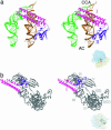
Stereo representation of relative binding position of RRF with respect to tRNAs and EF-G on the ribosome. (a) With respect to the A-(orange) and P-(green) site tRNAs. (b) With respect to EF-G (gray) in the GDP state. CCA, CCA-end side of the tRNAs; AC, anticodon side of the tRNAs; domains I and II of RRF and domains I–V of EF-G are denoted in respective matching colors. Orientations of the 70S ribosome in the two panels are represented by thumbnails at the lower right. In a, the 30S subunit is situated below the 50S subunit such that head of the 30S subunit and the CP of the 50S subunit face the viewer, whereas in b, the solvent side of the 30S subunit faces the viewer.

Similar articles

Cited by

References

    1. McCutcheon, J. P., Agrawal, R. K., Philips, S. M., Grassucci, R. A., Gerchman, S. E., Clemons, W. M., Jr., Ramakrishnan, V. & Frank, J. (1999) Proc. Natl. Acad. Sci. USA 96, 4301–4306. - PMC - PubMed
    1. Carter, A. P., Clemons, W. M., Jr., Brodersen, D. E., Morgan-Warren, R. J., Hartsch, T., Wimberly, B. T. & Ramakrishnan, V. (2001) Science 291, 498–501. - PubMed
    1. Stark, H., Rodnina, M. V., Rinke-Appel, J., Brimacombe, R., Wintermeyer, W. & van Heel, M. (1997) Nature 389, 403–406. - PubMed
    1. Agrawal, R. K., Penczek, P., Grassucci, R. A. & Frank, J. (1998) Proc. Natl. Acad. Sci. USA 95, 6134–6138. - PMC - PubMed
    1. Klaholz, B. P., Pape, T., Zavialov, A. V., Myasnikov, A. G., Orlova, E. V., Vestergaard, B., Ehrenberg, M. & van Heel, M. (2003) Nature 421, 90–94. - PubMed

Publication types

MeSH terms

LinkOut - more resources