. 2004 Jul;5(7):478-92.
doi: 10.1111/j.1398-9219.2004.00193.x.

Chemical genetic screening identifies sulfonamides that raise organellar pH and interfere with membrane traffic

Affiliations

Chemical genetic screening identifies sulfonamides that raise organellar pH and interfere with membrane traffic

Thomas J F Nieland et al. Traffic. 2004 Jul.

Abstract

Chemical genetics seeks to identify small molecules that afford functional dissection of cell biological pathways. Previous screens for small molecule inhibitors of exocytic membrane traffic yielded the identification and characterization of several compounds that block traffic from the Golgi to the cell surface as well as transport from the endoplasmic reticulum to the Golgi network [Feng et al. Proc Natl Acad Sci USA 2003;100:6469-6474; Yarrow et al. Comb Chem High Throughput Screen 2003;6:279-286; Feng et al. EMBO Reports 2004: in press]. Here, we screened these inhibitors for potential effects on endocytic membrane traffic. Two structurally related sulfonamides were found to be potent and reversible inhibitors of transferrin-mediated iron uptake. These inhibitors do not block endoplasmic reticulum-to-Golgi transport, but do disrupt Golgi-to-cell surface traffic. The compounds are members of a novel class of sulfonamides that elevate endosomal and lysosomal pH, down-regulate cell surface receptors, and impair recycling of internalized transferrin receptors to the plasma membrane. In vitro experiments revealed that the sulfonamides directly inhibit adenosine triphosphate (ATP) hydrolysis by the V-ATPase and that they also possess a potent proton ionophore activity. While maintenance of organellar pH is known to be a critical factor in both endocytosis and exocytosis, the precise role of acidification, beyond the uncoupling of ligands from their receptors, remains largely unknown. Identification of this novel class of sulfonamide inhibitors provides new chemical tools to better understand the function of organelle pH in membrane traffic and the activity of V-ATPases in particular.


Figures
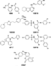
Figure 1
Figure 1
Chemical genetic screening identifies compounds that reduce exocytosis of GFP-tagged VSV-Gts045 to the cell surface. Structures of seven small mol-ecule compounds were identified in a screen of > 10000 chemicals from the Chembridge DIVERSet E library to block secretion of VSV G. Screening conducted by Y.F. (4,5).
Figure 2
Figure 2
Inhibition of VSV-Gts045 exocytosis. To examine the effects of chemicals shown in Figure 1, BSC1 cells were transduced with adenovirus to express GFP-tagged VSV Gts045 and grown overnight at 40 °C. Prior to transfer to 32 °C, 50 µm of each compound was added to the media for 1 h. After 3 h incubation at 32 °C, the amount of VSV Gts045 at the cell surface was measured by incubating with monoclonal VSV-G antisera as detailed in the Materials and Methods section. As a control for nonspecific binding, cells were also treated with 10µm brefeldin A, a drug known to completely block transport to the cell surface. After subtraction of background (measured in the presence of brefeldin A), data were normalized to the total fluorescence signal detected by GFP expression. Shown is the percentage of surface VSV-G relative to the total GFP signal (n=9; ±SEM). Experiments performed by Y.F.
Figure 3
Figure 3
Inhibitors of the exocytic pathway block(55)Fe uptake from Tf. HeLa cells were treated with 50 µm of inhibitors shown in Figure 1 for 4h at 37 °C (solid bars) or 4 °C (open bars) in the continued presence of 40nm 55FeTf. After washing, cells were lysed with 0.1% Triton X-100 containing 0.1% NaOH and cell-associated 55Fe was determined by liquid scintillation counting. Results were normalized to protein concentrations to determine pmol 55Fe/mg cell protein. The means of duplicate determinations are shown from a single experiment with similar results obtained on several occasions. Experiments were performed by J.X.B and P.D.B.
Figure 4
Figure 4
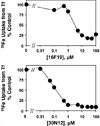
Dose-response to 16F19 and 30N12. Inhibition of 55Fe uptake from Tf was determined exactly as described for Figure 1. HeLa cells were incubated with the concentrations of inhibitors and the amount of 55Fe taken up by the cells was normalized to control cells which were treated with vehicle (DMSO) alone. Experiments performed by J.X.B and P.D.B.
Figure 5
Figure 5
16F19 and 30N12 inhibit 55Fe uptake from Tf in a reversible manner. HeLa cells were either first incubated with 2 µm 16F19 or 0.5 µm 30N12 for 1 h followed by a 4-h recovery incubation with 0.5% DMSO after removal of the drug, or cells were first incubated for 4 h with 0.5% DMSO followed by a 1 h incubation with sulfonamides. Cells treated under both conditions were then incubated with 40 nm 55Fe-Tf for 1h. During the 1-h assay, cells continued to be incubated either with DMSO (closed bars) to test reversibility (closed bars) or with sulfonamide to directly inhibitory potency in assay (open bars). 55Fe uptake was normalized to protein content and expressed as percentage control (cells incubated with 0.5% DMSO in the absence of sulfonamide). Experiments performed by P.D.B.
Figure 6
Figure 6
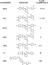
Family of sulfonamide inhibitors of membrane traffic. Based on the structures of 30N12 and 16D19, several additional compounds were screened for their ability to inhibit membrane traffic. Dose–response curves for inhibition of 55Fe uptake from Tf were measured as described for Figure 4. In addition to 30N12 and 16F19 (5), structures of 16L2, 16D2, 16D10, 23H9 and O-1 are shown along with the relative IC50 values determined for inhibition of Tf-mediated iron uptake. IC50 values were determined as described in Materials and Methods using GraphPad PRISM3 software. Experiments performed by J.X.B and P.D.B. with data analysis by T.J.F.N.
Figure 7
Figure 7
16D10 sulfonamide does not inhibit NTBI uptake. The uptake of 55Fe was measured in the absence of Tf by using a chemical chelate with NTA (1: 4 ratio) to present the cation to HeLa cells. To measure nontransferrin bound iron (NTBI) uptake, cells were preincubated with 50µm of the indicated compounds for 30 min prior to addition of 2µm 55FeNTA. After 1 h incubation at either 4 °C (open bars) or 37 °C (closed bars), uptake was quenched by placing the cells on ice and incubating with unlabeled 1 mm FeNTA to displace surface-bound iron; lysates were collected to measure cell-associated radioactivity and protein to calculate pmol 55Fe/mg cell protein. Shown are the means of duplicate determinations from a single experiment with similar results obtained on several occasions. Experiments performed by J.X.B and P.D.B.
Figure 8
Figure 8
Sulfonamides block VSV-Gts045 transport from the Golgi apparatus to the plasma membrane. Cells were transduced to express VSV-Gts045 and incubated as described for Figure 2 except that 100µm of 16D10 or O-1 were added before transfer to 32 °C. After 30-min and 2-h incubation periods, the cells were fixed with 3% formaldehyde and images of GFP-tagged VSV-Gts045 were collected using a 40× lens. Scale bar = 10µm. Experiments performed by Y.F.
Figure 9
Figure 9
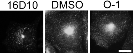
16D10 sulfonamide reduces entry of Tf into cells. TRVb-1 cells were pretreated with 10µm 16D10, O-1 or DMSO for 30 min and then 40µg/mL Alexis594-labeled Tf was added for an additional 30 min. Cells were fixed with 3% formaldehyde and fluorescence images were collected using the same exposure times with a 63× oil immersion lens. Scale bar = 5 µm. Experiments performed by T.J.F.N.
Figure 10
Figure 10
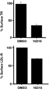
16D10 sulfonamide promotes down-regulation of cell surface receptors. Top panel: TRVb-1 cells were preincubated in serum free media for 90min at 37 °C, then treated with or without 10µm 16D10 for 30min at 37 °C. After cells were chilled on ice, surface 125I-Tf binding was measured as described under Materials and Methods. Data shown are the average (± SD) of two experiments. The data are plotted as a fraction of the 125I-Tf bound to the surface of control cells. Bottom panel: TVRb-1 cells were grown for 3 days in medium supplemented with lipoprotein deficient serum to up-regulate endogenous LDL receptor. On the day of assay, cells were washed twice in serum-free medium and pretreated with 10µm 16D10 or DMSO alone for 30 min at 37 °C. Cells were then chilled on ice, and LDL labeled with 125I on the lipoprotein moiety was added for 1 h on ice at a final concentration of 25µg/mL. Non-bound LDL was washed away with phosphate-buffered saline and cells were lysed in 0.1N NaOH for 30 min at room temperature. For each sample, an aliquot was counted by LSC and assayed for protein levels. Data are expressed as LDL receptor levels as a percentage of control. Note that saturating levels of ligands were used to measure cell surface binding for Tf and LDL receptors, 3µg/mL and 25µg/mL, respectively. Experiments performed by T.J.F.N and T.D.C.
Figure 11
Figure 11
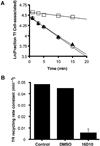
16D10 sulfonamide impairs Tf receptor recycling. Panel A: TRVb-1 cells (treated with or without 16D10) were incubated with 125I-Tf for 2h at 37 °C to allow ligand internalization, then washed and incubated for the indicated time to allow recycling and release of Tf. The amount of 125I-Tf remaining as a function of time is shown for control (open circles), cells treated with 0.5% DMSO (closed triangles) and cells treated with 10µm 16D10 (open squares). Panel B: Recycling rate constant (min −1) was determined from the slope of lines shown in the middle panel; shown is the mean value (±SD) determined in four separate experiments. Experiments performed by T.D.C.
Figure 12
Figure 12
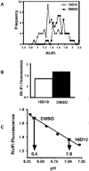
16D10 sulfonamide alters endosomal pH. Panel A: Histogram of the distribution of Rh/Fl ratio of peri-centriolar recycling compartments of control and 16D10 treated cells. Compound 16D10 causes an increase in the Fl fluorescence, resulting in a shift of the distribution to the left. The data are from a representative experiment and 60 peri-centriolar recycling compartments from each condition were measured. Panel B: The average ± SEM Rh/Fl ratio of peri-centriolar recycling compartment from control and 16D10 treated cells (n = 60). Panel C: Estimation of endosomal pH based on the Rh/Fl ratio. A standard pH curve was constructed by determining the Rh/Fl ratio in cells whose endosomes were equilibrated to a known pH (squares). The Rh/Fl ratio measured in control and 16D10 cells can be converted to pH using this standard curve. The peri-centriolar endosomes in controls cells have a pH of ~6.4 and those in cells treated with compound 16D10 have a pH of ~7.0. Experiments performed by T.D.C.
Figure 13
Figure 13
16D10 and 16D2 prevent lysosomal degradation. K562 cells were incubated with β-galactosidase at 20 °C for 60 min to allow its internalization by fluid phase into endosomal (prelysosomal) compartments. After washing, cells were incubated for 30min in the presence of 10µm 16D10 or 1% DMSO vehicle control, 0.5µm bafilomycin A1 (BafA1) or 20 mm NH4Cl. The intracellular activity of internalized β-galactosidase following the chase period reflects access to lysosomes as is manifested by decreased β-galactosidase activity due to its proteolytic degradation within lysosomes. Results of duplicate samples were normalized to the total amount of activity before the chase period and are expressed as percentage control (DMSO vehicle treatment). Experiment performed by T.J.F.N.
Figure 14
Figure 14
Proton ionophore activity of sulfonamides. Panel A: Bovine brain V-ATPase was reconstituted into proteoliposomes and assayed for ATP-driven proton transport activity. Sulfonamide analog 16D2 (10 nm) added at the end of the assay was found to collapse the proton gradient. Panel B: The proton ionophore activity of sulfonamides was measured as membrane potential-driven proton conductance in a protein-free liposome system. Sulfonamides and 1799 were added at the indicated concentrations. Experiments performed by J.W.

Similar articles

Cited by

References

    1. Specht KM, Shokat KM. The emerging power of chemical genetics. Curr Opin Cell Biol. 2002;14:155–159. - PubMed
    1. Stockwell BR. Frontiers in chemical genetics. Trends Biotechnol. 2000;18:449–455. - PubMed
    1. Stockwell BR. Chemical genetics: ligand-based discovery of gene function. Nat Rev Genet. 2000;1:116–125. - PMC - PubMed
    1. Feng Y, Yu S, Lasell TK, Jadhav AP, Macia E, Chardin P, Melancon P, Roth M, Mitchison T, Kirchhausen T. Exo1: a new chemical inhibitor of the exocytic pathway. Proc Natl Acad Sci U S A. 2003;100:6469–6474. - PMC - PubMed
    1. Yarrow JC, Feng Y, Perlman ZE, Kirchhausen T, Mitchison TJ. Phenotypic screening of small molecule libraries by high throughput cell imaging. Comb Chem High Throughput Screen. 2003;6:279–286. - PubMed

Publication types

MeSH terms