

Changes in contractile activation characteristics of rat fast and slow skeletal muscle fibres during regeneration
- PMID: 15181161
- PMCID: PMC1664957
- DOI: 10.1113/jphysiol.2004.066217
Changes in contractile activation characteristics of rat fast and slow skeletal muscle fibres during regeneration
Abstract
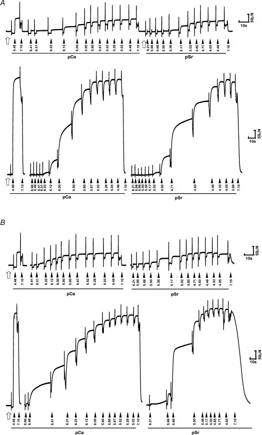
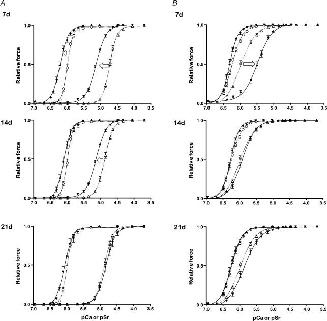
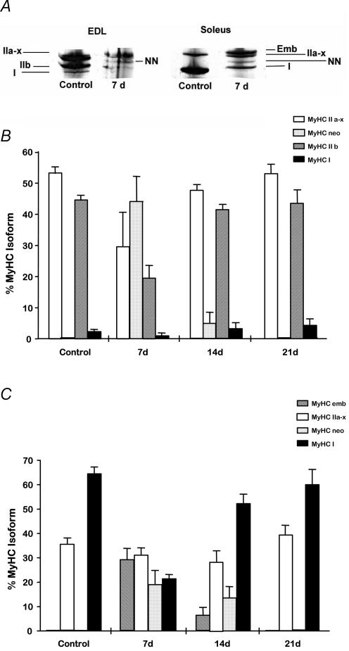
Damaged skeletal muscle fibres are replaced with new contractile units via muscle regeneration. Regenerating muscle fibres synthesize functionally distinct isoforms of contractile and regulatory proteins but little is known of their functional properties during the regeneration process. An advantage of utilizing single muscle fibre preparations is that assessment of their function is based on the overall characteristics of the contractile apparatus and regulatory system and as such, these preparations are sensitive in revealing not only coarse, but also subtle functional differences between muscle fibres. We examined the Ca(2+)- and Sr(2+)-activated contractile characteristics of permeabilized fibres from rat fast-twitch (extensor digitorum longus) and slow-twitch (soleus) muscles at 7, 14 and 21 days following myotoxic injury, to test the hypothesis that fibres from regenerating fast and slow muscles have different functional characteristics to fibres from uninjured muscles. Regenerating muscle fibres had approximately 10% of the maximal force producing capacity (P(o)) of control (uninjured) fibres, and an altered sensitivity to Ca(2+) and Sr(2+) at 7 days post-injury. Increased force production and a shift in Ca(2+) sensitivity consistent with fibre maturation were observed during regeneration such that P(o) was restored to 36-45% of that in control fibres by 21 days, and sensitivity to Ca(2+) and Sr(2+) was similar to that of control (uninjured) fibres. The findings support the hypothesis that regenerating muscle fibres have different contractile activation characteristics compared with mature fibres, and that they adopt properties of mature fast- or slow-twitch muscle fibres in a progressive manner as the regeneration process is completed.
Figures
Similar articles
-
Matching of sarcoplasmic reticulum and contractile properties in rat fast- and slow-twitch muscle fibres.Clin Exp Pharmacol Physiol. 2006 Jul;33(7):591-600. doi: 10.1111/j.1440-1681.2006.04412.x. Clin Exp Pharmacol Physiol. 2006. PMID: 16789925
-
Dependence of cross-bridge kinetics on myosin light chain isoforms in rabbit and rat skeletal muscle fibres.J Physiol. 2006 Feb 15;571(Pt 1):231-42. doi: 10.1113/jphysiol.2005.099770. Epub 2005 Dec 15. J Physiol. 2006. PMID: 16357018 Free PMC article.
-
Potential targets for skeletal muscle impairment by hypogravity: basic characterization of resting ionic conductances and mechanical threshold of rat fast- and slow-twitch muscle fibers.J Gravit Physiol. 1998 Jul;5(1):P75-6. J Gravit Physiol. 1998. PMID: 11542372
-
Events of the excitation-contraction-relaxation (E-C-R) cycle in fast- and slow-twitch mammalian muscle fibres relevant to muscle fatigue.Acta Physiol Scand. 1998 Mar;162(3):229-45. doi: 10.1046/j.1365-201X.1998.0304f.x. Acta Physiol Scand. 1998. PMID: 9578368 Review.
-
The effect of age on in vitro motility speed of slow myosin extracted from single rat soleus fibres.Acta Physiol Scand. 1999 Dec;167(4):325-6. doi: 10.1046/j.1365-201x.1999.00619.x. Acta Physiol Scand. 1999. PMID: 10632634 Review.
Cited by
-
Structural and functional alterations of muscle fibres in the novel mouse model of facioscapulohumeral muscular dystrophy.J Physiol. 2007 Nov 1;584(Pt 3):997-1009. doi: 10.1113/jphysiol.2007.141481. Epub 2007 Sep 13. J Physiol. 2007. PMID: 17855756 Free PMC article.
-
Shrimp Protein Hydrolysate Modulates the Timing of Proinflammatory Macrophages in Bupivacaine-Injured Skeletal Muscles in Rats.Biomed Res Int. 2016;2016:5214561. doi: 10.1155/2016/5214561. Epub 2016 Oct 27. Biomed Res Int. 2016. PMID: 27868064 Free PMC article.
-
Ageing prolongs inflammatory marker expression in regenerating rat skeletal muscles after injury.J Inflamm (Lond). 2011 Dec 29;8(1):41. doi: 10.1186/1476-9255-8-41. J Inflamm (Lond). 2011. PMID: 22206492 Free PMC article.
-
Beneficial effects of cod protein on inflammatory cell accumulation in rat skeletal muscle after injury are driven by its high levels of arginine, glycine, taurine and lysine.PLoS One. 2013 Oct 4;8(10):e77274. doi: 10.1371/journal.pone.0077274. eCollection 2013. PLoS One. 2013. PMID: 24124612 Free PMC article.
-
Impact of pregnancy and vaginal delivery on the passive and active mechanics of the rat vagina.Ann Biomed Eng. 2011 Jan;39(1):549-58. doi: 10.1007/s10439-010-0153-9. Epub 2010 Sep 8. Ann Biomed Eng. 2011. PMID: 20824342 Free PMC article.
References
-
- Bigard XA, Janmot C, Merino D, Lienhard F, Guezennec YC, d'Albis A. Endurance training affects myosin heavy chain phenotype in regenerating fast-twitch muscle. J Appl Physiol. 1996;81:2658–2665. - PubMed
-
- Bigard AX, Zoll J, Ribera F, Mateo P, Sanchez H, Serrurier B, Ventura-Clapier R. Influence of overload on phenotypic remodeling in regenerated skeletal muscle. Am J Physiol. 2001;281:C1686–C1694. - PubMed
-
- Bortolotto SK, Cellini M, Stephenson DG, Stephenson GM. MHC isoform composition and Ca2+- or Sr2+-activation properties of rat skeletal muscle fibers. Am J Physiol. 2000;279:C1564–C1577. - PubMed
-
- Bottinelli R. Functional heterogeneity of mammalian single muscle fibres: do myosin isoforms tell the whole story? Pflugers Arch. 2001;443:6–17. - PubMed
-
- Bottinelli R, Reggiani C. Human skeletal muscle fibres: molecular and functional diversity. Prog Biophys Mol Biol. 2000;73:195–262. - PubMed
Publication types
MeSH terms
Substances
LinkOut - more resources
Full Text Sources
Research Materials
Miscellaneous