. 2004 Jun 15;101(24):9103-8.
doi: 10.1073/pnas.0402959101. Epub 2004 Jun 4.

The Parkinson's disease protein DJ-1 is neuroprotective due to cysteine-sulfinic acid-driven mitochondrial localization

Affiliations

The Parkinson's disease protein DJ-1 is neuroprotective due to cysteine-sulfinic acid-driven mitochondrial localization

Rosa M Canet-Avilés et al. Proc Natl Acad Sci U S A. .

Abstract

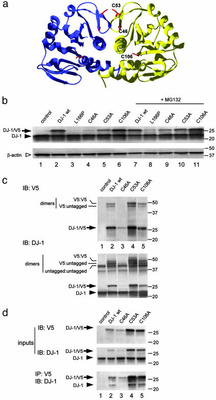
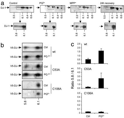
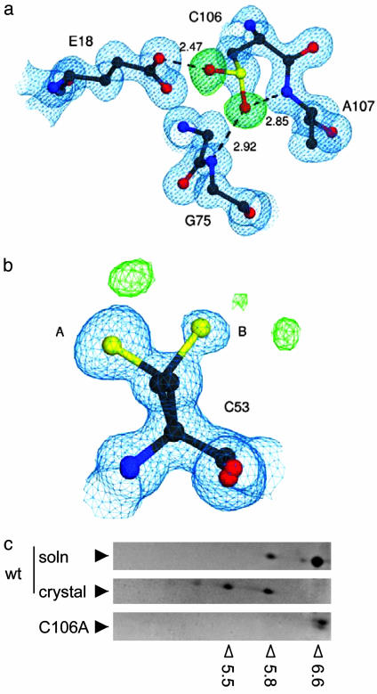
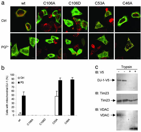
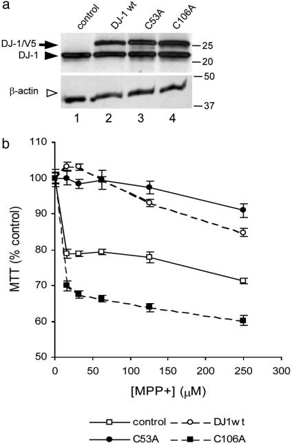
Loss-of-function DJ-1 mutations can cause early-onset Parkinson's disease. The function of DJ-1 is unknown, but an acidic isoform accumulates after oxidative stress, leading to the suggestion that DJ-1 is protective under these conditions. We addressed whether this represents a posttranslational modification at cysteine residues by systematically mutating cysteine residues in human DJ-1. WT or C53A DJ-1 was readily oxidized in cultured cells, generating a pI 5.8 isoform, but an artificial C106A mutant was not. We observed a cysteine-sulfinic acid at C106 in crystalline DJ-1 but no modification of C53 or C46. Oxidation of DJ-1 was promoted by the crystallization procedure. In addition, oxidation-induced mitochondrial relocalization of DJ-1 and protection against cell death were abrogated in C106A but not C53A or C46A. We suggest that DJ-1 protects against neuronal death, and that this is signaled by acidification of the key cysteine residue, C106.


Figures

Fig. 1.
Fig. 1.
Generation of stable dimeric cysteine mutants of human DJ-1. (a) The crystal structure of human DJ-1 with cysteine residues highlighted. Two cysteine residues, C46 and C53, whose sidechains are shown in red, are present in a β-turn close to the dimer interface. A third cysteine, C106, is within the center of the dimer. (b) Transient transfections of V5-tagged cysteine mutants into M17 cells. Because some variants, notably C46A, were unstable, samples in lanes 7-11 were treated with 5 μM MG132 for 24 h. Western blots were probed with anti-DJ-1 (Upper) to visualize both transfected DJ-1 (arrows) and endogenous DJ-1 (arrowhead). The blot was reprobed with β-actin as a loading control (Lower, open arrowhead). Markers on the right of the blot are in kilodaltons. (c) To assess whether these variants form dimers, transfected cells were treated with the crosslinker disuccinimidyl suberate before extraction and Western blotting for V5 (Upper) or DJ-1 (Lower). Monomeric V5-tagged protein is indicated with an arrow, and endogenous DJ-1 in the cells indicated by an arrowhead. The V5-tagged proteins formed dimers with either endogenous or transfected DJ-1, indicated by lines on the left of the blot. Blots are representative of at least duplicate experiments. To confirm these results, we coimmunoprecipitated V5-tagged DJ-1 with endogenous protein (d). (Upper) Lysates of transfected cells probed with V5 or DJ-1 to demonstrate input levels of proteins. (Lower) Lysates immunoprecipitated withV5 antibody and blotted with DJ-1 antibody to show interaction of endogenous DJ-1 with the tagged proteins.
Fig. 2.
Fig. 2.
Oxidation of DJ-1 is prevented in the C106A mutant. (a) Endogenous DJ-1 in M17 cells responds to oxidative stress. (Upper) From left to right, blots of 2D gels from extracts from untreated cells, cells exposed to PQ2+ or MPP+; on the right, from cells exposed to PQ2+ for 24 h then placed in fresh media for 24 h. Blots were probed with monoclonal anti-DJ1 (arrow), which shows multiple isoforms: numbers below the blots show approximate pI values for each of the major isoforms. (Inset) Area around the pI 5.8 isoform, which accumulates after oxidative stress. (b) Response to oxidative stress in cysteine mutants. Samples were extracted from cells transfected with V5-tagged WT, C53A, or C106A DJ-1, from either untreated cells or cells exposed to PQ2+. Blots were probed with monoclonal anti-V5 and the pI 5.8 isoform identified (indicated below). The pI 5.8 isoform accumulated with both WT and C53A variants but not with the C106A variant (Bottom). Quantification of this and three similar experiments in c shows the ratio of pI 5.8:pI 6.1 isoform (n = 4; bars represent SEM). Note that the y axis scales are different for C106A; we included the faint immunoreactivity in the pI 5.8 region, but the ratio was still substantially lower than for the other variants.
Fig. 3.
Fig. 3.
Identification of C106-SO2H in the crystal. (a) Immediate environment of C106-SO2H, with 2mFO-DFC electron density contoured at 1.0 σ (blue) and mFO-DFC electron density contoured at 4.0 σ (green) m and D are weights for the Fourier coefficients. Both electron density maps were calculated before the introduction of the Oδ1 and Oδ2 oxygen atoms of C106-SO2H. Dotted lines indicate hydrogen bonds between C106-SO2H and the surrounding residues E18, G75, and A107, with lengths in angstroms. (b) C53 is not modified to the same extent as C106, although this residue can adopt two conformations (A and B). Difference electron densities (green) represent probable minor modifications. This figure was made with povscript+ (28). (c) Accumulation of acidic pI isoforms after crystallization. Recombinant WT DJ-1 before (Top) or after crystallization (Middle) were separated on 2D gels and different isoforms identified by Coomassie staining. Note that the crystallization process induces a pI shift in the recombinant protein, although a basal level of oxidized protein is seen in solution. In contrast, C106A is present only as the most basic isoform (pI 6.6-6.8). Open arrowheads below the gels show experimentally determined pI values, which were calibrated with 2D gel standards; filled arrowheads indicate the position of DJ-1.
Fig. 4.
Fig. 4.
DJ-1 localizes to the outer mitochondrial membrane after oxidative stress. (a) M17 cells were transfected with V5-tagged DJ-1 variants as indicated and stained for DJ-1 (V5, green) and mitotracker (red). After exposure to PQ2+ (Lower), DJ-1 colocalized with mitochondria much more extensively than under control conditions (Upper), as evidenced by the yellow color where staining overlaps. The C106A and C106D variants did not relocalize to mitochondria, whereas C53A and C46A did. This finding is quantified in b by counting the proportion of V5-positive cells that showed strong colocalization with mitotracker. Representative data are shown from at least three experiments for each construct, performed in duplicate. (c) DJ-1 is not imported into mitochondria. Cells transfected with WT DJ-1 were exposed to PQ2+ and mitochondrial fractions prepared. Duplicate samples were either untreated (-, lanes 1 and 2) or subjected to limited proteolysis with trypsin (+, lanes 3 and 4) and blotted for V5 (Top), the outer mitochondrial membrane protein VDAC1 (Middle), or the inner mitochondrial membrane protein Tim23 (Bottom).
Fig. 5.
Fig. 5.
Cysteine mutants affect the ability of DJ-1 to protect against mitochondrial damage. For toxicity assays, we generated stable cell lines that expressed either WT, C53A, or C106A DJ-1 (a). Western blots show expression of the V5-tagged constructs (arrow) and endogenous DJ-1 (filled arrowhead) and were reprobed with β-actin (Lower, open arrowhead). (b) Exposure to MPP+ for 48 h resulted in cell death, assessed using 3-(4,5-dimethylthiazol-2-yl)-2,5-diphenyl tetrazolium bromide (MTT) assays. Cells expressing WT or C53A DJ-1 were resistant to MPP+ toxicity, whereas cells expressing C106A DJ-1 showed increased sensitivity to this toxin. Data are shown from one set of experiments with all four cell lines performed on the same day, expressed as a percentage of untreated cells (n = 8 wells per dose, error bars indicate SEM) and are representative of at least triplicate experiments per cell line.

References

    1. Dawson, T. M. & Dawson, V. L. (2003) Science 302, 819-822. - PubMed
    1. Petrucelli, L., O'Farrell, C., Lockhart, P. J., Baptista, M., Kehoe, K., Vink, L., Choi, P., Wolozin, B., Farrer, M., Hardy, J., et al. (2002) Neuron 36, 1007-1019. - PubMed
    1. Darios, F., Corti, O., Lucking, C. B., Hampe, C., Muriel, M. P., Abbas, N., Gu, W. J., Hirsch, E. C., Rooney, T., Ruberg, M., et al. (2003) Hum. Mol. Genet. 12, 517-526. - PubMed
    1. Bonifati, V., Rizzu, P., van Baren, M. J., Schaap, O., Breedveld, G. J., Krieger, E., Dekker, M. C., Squitieri, F., Ibanez, P., Joosse, M., et al. (2003) Science 299, 256-259. - PubMed
    1. Taira, T., Saito, Y., Niki, T., Iguchi-Ariga, S. M., Takahashi, K. & Ariga, H. (2004) EMBO Rep. 5, 213-218. - PMC - PubMed

Publication types

MeSH terms

Associated data