

doi: 10.1042/BJ20040846.
PKA-phosphorylation of PDE4D3 facilitates recruitment of the mAKAP signalling complex
Affiliations
- PMID: 15182229
- PMCID: PMC1133866
- DOI: 10.1042/BJ20040846
Item in Clipboard
PKA-phosphorylation of PDE4D3 facilitates recruitment of the mAKAP signalling complex
Biochem J.
.
Abstract
mAKAP (muscle-selective A-kinase-anchoring protein) co-ordinates a cAMP-sensitive negative-feedback loop comprising PKA (cAMP-dependent protein kinase) and the cAMP-selective PDE4D3 (phosphodiesterase 4D3). In vitro and cellular experiments demonstrate that PKA-phosphorylation of PDE4D3 on Ser-13 increases the affinity of PDE4D3 for mAKAP. Our data suggest that activation of mAKAP-anchored PKA enhances the recruitment of PDE4D3, allowing for quicker signal termination.
Figures
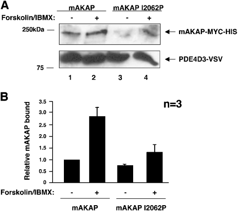
(A) Wild-type mAKAP (lanes 1 and 2) and a mutant form of mAKAP that cannot anchor PKA (mAKAP I2062P) (lanes 3 and 4) were transfected into HEK-293 cells with PDE4D3–VSV, as indicated. At 2 days after transfection, cells were serum-starved for 5 h and stimulated with 10 μM forskolin and 50 μM IBMX for 15 min, as indicated. PDE4D3–VSV protein was immunoprecipitated with anti-VSV antibody, resolved by SDS/7% PAGE and transferred on to nitrocellulose. Immunoprecipitates were probed with anti-mAKAP–HRP to detect mAKAP (upper panel). Anti-VSV antibody was used to detect PDE4D3–VSV to show equivalent levels of immunoprecipitated PDE4D3 (lower panel). These blots are representative of three independent experiments. (B) Bands from (A) were quantified using NIH Image software. The amount of mAKAP bound to PDE4D3–VSV was normalized to the amount of PDE4D3–VSV immunoprecipitated. Results are means±S.E.M. for three independent experiments. The columns are labelled with transfection and stimulation conditions.
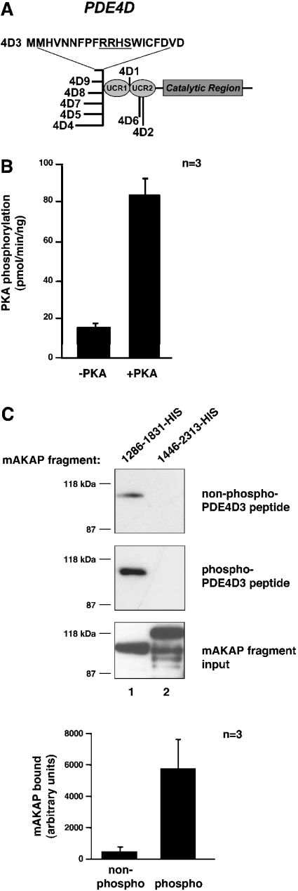
(A) The structure of the PDE4D family of PDEs. The conserved catalytic core and upstream conserved regions (UCRs) are indicated. The locations of divergent sequences for each isoform are indicated, and the 20 unique amino acid residues of PDE4D3 are denoted by the one-letter amino acid code. The PKA-phosphorylation consensus site is underlined. (B) Phosphorylation of PDE4D3 peptide by PKA. PDE4D3 peptide was incubated in PKA kinase buffer containing 100 μM ATP, 5 μM [γ-32P]ATP and 0.3 mM cAMP with or without the PKA catalytic subunit for 15 min at 30 °C. The reaction mixture was spotted on to p81 phosphocellulose paper, washed five times in 75 mM phosphoric acid and once in 95% ethanol. Filters were air-dried and were counted by liquid-scintillation counting. Results are averages of three independent experiments. (C) Pull-down assay of mAKAP fragments with PDE4D3 peptide. Biotin–PDE4D3 peptide was incubated in PKA kinase buffer containing 100 μM ATP and 0.3 mM cAMP with or without the PKA catalytic subunit for 2 h at 30 °C. Neutravidin beads were added to collect the peptide and then rocked for 1 h at room temperature. The precipitates were washed three times and resuspended in 500 μl of HSE buffer containing 5 μM recombinant mAKAP fragments. Reactions were incubated overnight at 4 °C with rocking. Precipitates were washed three times with HSE buffer, and bound proteins were subjected to SDS/7% PAGE followed by immunoblotting with anti-His antibody. The top blot tests co-precipitation of mAKAP fragments with nonphosphorylated PDE4D3 peptide. The middle blot tests co-precipitation of mAKAP fragments with phosphorylated PDE4D3 peptide. The bottom blot shows that equal amounts of mAKAP fragments (lane 1, mAKAP 1286–1831–His; lane 2, mAKAP 1446–2313–His) were used in the assay. The 1286–1831–His bands from the top and middle blots were quantified using NIH Image software. The amount of mAKAP fragment bound to non-phosphorylated (non-phospho) and PKA-phosphorylated (phospho) peptides was graphed in arbitrary units. Results are means±S.E.M. for three independent experiments.
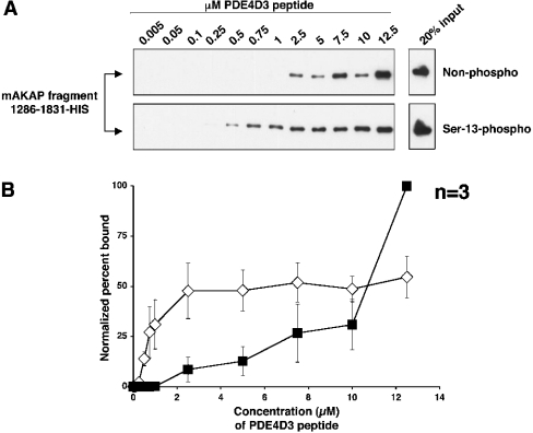
(A) Biotin–PDE4D3 peptides that were either non-phosphorylated (upper panel) or phosphorylated on Ser-13 (lower panel) were mixed at various concentrations (indicated in μM) with a fixed amount of mAKAP fragment 1286–1831–His. Biotin–PDE4D3 peptides were collected by Neutravidin beads, resolved by SDS/7% PAGE and subjected to Western analysis using anti-His antibody to detect the mAKAP fragment. The upper and lower right-hand panels represent 20% of the mAKAP fragment added to the samples. The blots shown are representative of three independent experiments. (B) Bands from (A) were quantified using NIH Image for each experiment, and the percentage of mAKAP 1286–1831–His bound to peptide for each concentration point was determined (see the Experimental section). To create the graph, the normalized percentage of mAKAP 1286–1831–His bound to biotin–Ser-13-phosphorylated PDE4D3 peptide (⋄) or biotin–non-phosphorylated PDE4D3 peptide (▪) was plotted against peptide concentration. Results are the means±S.E.M. for three independent experiments.
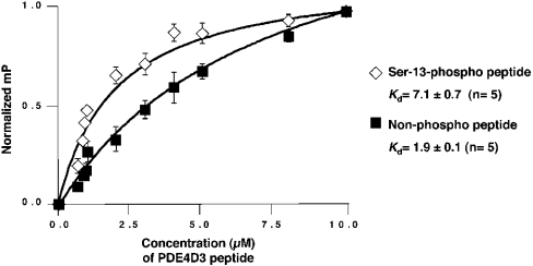
FITC–Ser-13-phosphorylated PDE4D3 peptide (⋄) or FITC–non-phosphorylated PDE4D3 peptide (▪) was resuspended in PBS containing 5 mg/ml BSA, pH 7.0. Increasing concentrations of recombinant mAKAP 1286–1831–His were mixed with each FITC-labelled peptide. Each sample was incubated for 10 min. Saturation-binding curves were generated with Prism graphing software. Milli-polarization (mP)=FP/1000. Kds were calculated from non-linear regression curves generated from the average of five individual experiments.
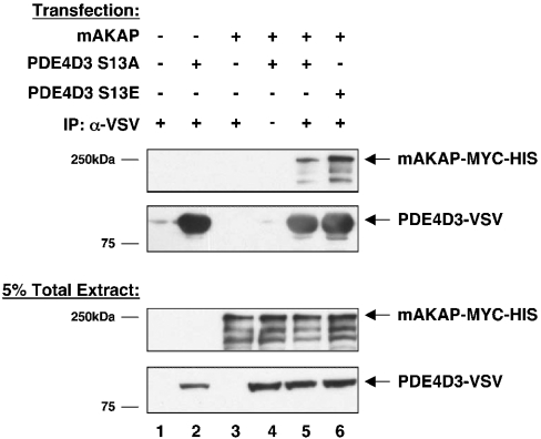
PDE4D3–VSV phospho-mutants were created to mimic the non-phosphorylated form (PDE4D3–VSV Ser13Ala) or the phosphorylated form (PDE4D3–VSV Ser13Glu). PDE4D3–VSV phosphomutants were co-transfected with mAKAP into HEK-293 cells as indicated. At 2 days after transfection, PDE4D3–VSV protein was immunoprecipitated with anti-VSV antibody. Immunoprecipitates were probed with anti-mAKAP–HRP to detect mAKAP (top panel) and anti-VSV to detect PDE4D3 phospho-mutants (second panel). The input (5% of total extract) was immunoblotted with anti-mAKAP–HRP (third panel) or anti-VSV (bottom panel) antibodies to demonstrate equivalent expression levels of mAKAP and PDE4D3 phospho-mutants in all transfection conditions. These blots are representative of five independent experiments.
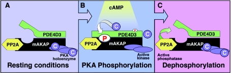
(A) Under resting conditions, PDE4D3 binds to the mAKAP signalling complex. (B) Upon hormonal stimulation of a cell, cAMP increases and activates mAKAP-bound PKA. The catalytic subunits phosphorylate local substrates, including PDE4D3. Phosphorylation of PDE4D3 on Ser-13 enhances the binding affinity of PDE4D3 for mAKAP. Phosphorylation of PDE4D3 on Ser-54 increases PDE activity 2-fold and causes cAMP breakdown. (C) With the return of basal cAMP levels, the PKA holoenzyme reforms. Phosphatase activity dephosphorylates PDE4D3, and the signalling system is reset.
References
-
- Colledge M., Scott J. D. AKAPs: from structure to function. Trends Cell Biol. 1999;9:216–221. - PubMed
-
- Tasken K., Aandahl E. M. Localized effects of cAMP mediated by distinct routes of protein kinase A. Physiol. Rev. 2004;84:137–167. - PubMed
-
- Francis S. H., Turko I. V., Corbin J. D. Cyclic nucleotide phosphodiesterases: relating structure and function. Prog. Nucleic Acid Res. Mol. Biol. 2001;65:1–52. - PubMed
-
- Yarwood S. J., Steele M. R., Scotland G., Houslay M. D., Bolger G. B. The RACK1 signaling scaffold protein selectively interacts with the cAMP-specific phosphodiesterase PDE4D5 isoform. J. Biol. Chem. 1999;274:14909–14917. - PubMed
Publication types
MeSH terms
Substances
Grants and funding
LinkOut - more resources
Full Text Sources
Other Literature Sources
Molecular Biology Databases