. 2004 Jun;70(6):3609-17.
doi: 10.1128/AEM.70.6.3609-3617.2004.

Unusual microbial xylanases from insect guts

Affiliations

Unusual microbial xylanases from insect guts

YaLi Brennan et al. Appl Environ Microbiol. 2004 Jun.

Abstract

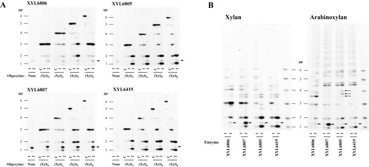
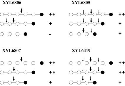
Recombinant DNA technologies enable the direct isolation and expression of novel genes from biotopes containing complex consortia of uncultured microorganisms. In this study, genomic libraries were constructed from microbial DNA isolated from insect intestinal tracts from the orders Isoptera (termites) and Lepidoptera (moths). Using a targeted functional assay, these environmental DNA libraries were screened for genes that encode proteins with xylanase activity. Several novel xylanase enzymes with unusual primary sequences and novel domains of unknown function were discovered. Phylogenetic analysis demonstrated remarkable distance between the sequences of these enzymes and other known xylanases. Biochemical analysis confirmed that these enzymes are true xylanases, which catalyze the hydrolysis of a variety of substituted beta-1,4-linked xylose oligomeric and polymeric substrates and produce unique hydrolysis products. From detailed polyacrylamide carbohydrate electrophoresis analysis of substrate cleavage patterns, the xylan polymer binding sites of these enzymes are proposed.


Figures

FIG. 1.
FIG. 1.
Schematic diagrams of xylanase primary sequences isolated from insect gut microbial libraries. (A) XYL6806 is a very unique xylanase belonging to family 8. It has a large N-terminal domain that returns no BLAST hits against the National Center for Biotechnology Information database. It is linked to the catalytic domain with a proline-rich linker and has a C-terminal carbohydrate-binding module. (B) Schematic diagrams of the domain structures of three family 11 xylanases. All three are very unique enzymes and have very low identity to known xylanases but clearly contain all family 11 signature motifs and catalytic residues. XYL6807 has a C-terminal domain that returns no BLAST hits against the National Center for Biotechnology Information database.
FIG. 2.
FIG. 2.
Sequence alignments showing the conserved regions in the family 8 (XYL6806) and family 11 (XYL6807, XYL6419, and XYL6805) xylanases discovered from insect gut samples. Regions of significant sequence identity or similarity are shown in boldface. Catalytic residues are shown with white text on black, and residues known to be involved in substrate binding are boxed. All published family 8 sequences (family 8 includes glucanase, chitosanase, and xylanase sequences) were used to construct the family 8 alignment. However, for illustration purposes, only the xylanase sequences are shown (B. hal., Bacillus halodurans; B. sp., Bacillus sp. strain KK-1; P. hal, Pseudoalteromonas haloplanktis). For the family 11 alignment, all published family 11 xylanases were used, and the most similar sequences to XYL6419, XYL6807, and XYL6805 are shown (P. co., Piromyces communis; N. fr., Neocallimastix frontalis; O. sp, Orpinomyces sp. strain PC-2; R. al., Ruminococcus albus) as well as some selected more common xylanases (A. ni, Aspergillus niger; T. re., Trichoderma reesei; B. st., Bacillus stearothermophilus; S. co., Streptomyces coelicolor).
FIG. 3.
FIG. 3.
Phylogenetic tree of family 11 catalytic domain sequences generated by the neighbor-joining method. At major nodes, bootstrap values for 1,000 resamplings are shown. One Firmicutes group contains organisms of the orders Bacillales and Clostridiales (GenBank accession no. U76545, X00660, AF326785, AF220528, X59059, AB010958, Z11127, U43089, AJ272430, AJ132472, Z49970, AF036925, M31726, and D13325) and two sequences from the ciliate P. multivesiculatum (accession no. AJ009828 and AB011274). The other Firmicutes group contains only sequences from the order Bacillales (GenBank accession no. U15985, X59059, AP001510, U51675, AF195421, and D32065) and are branched with the yeast xylanase from Pichia stipitis (AF151379). All of the actinobacterial xylanase sequences form a clade (GenBank accession no. X76729, AF120156, X81045, X98518, AJ292317, AL109949, AL133220, AF198618, AF194025, and U01242). The fungal xylanases form a large group containing the phyla Ascomycota and Basidiomycota (GenBank accession no. AJ238895, X76047, U35436, AF169630, U58915, U58916, L13596, AB003085, U39784, S45138, L26988, O43097, AJ278385, Z50050, AB035540, Z68891, Y16969, X69573, X69574, AF246831, AF246830, P48793, AJ012718, D49850, D49851, L37529, Z81317, A44597, AF301904, and D63382) and two bacterial xylanase sequences (Pseudomonas fluorescens [Z48927] and Cellvibrio mixtus [Z48925]) and the xylanase from the beetle Phaedon cochleariae (Y17908). The three insect gut xylanases discovered in this study (XYL6807, XYL6805, and XYL6419) branch with xylanases from the rumen fungi Neocallimastix frontalis (X82439), Orpinomyces sp. strain PC-2 (U57819), and Piromonas communis (AF297649) and the rumen bacterium Ruminococcus albus (AB057588).
FIG. 4.
FIG. 4.
Maximum-likelihood tree of all family 8 catalytic domain sequences. Numbers at nodes are bootstrap values for 100 resamplings. The GenBank accession numbers for the sequences used to calculate the tree are shown in parentheses. The tree is formed of two endoglucanase clades: a xylanase clade containing XYL6806 and a clade consisting of chitosanase sequences.
FIG. 5.
FIG. 5.
Hydrolysis product fingerprints of xylanases XYL6806, XYL6807, XYL6805, and XYL6419. DP, degree of polymerization. (A) Oligosaccharides obtained after hydrolysis of oligoxylans (Xyl)3, (Xyl)4, (Xyl)5, and (Xyl)6. All reactions were performed at pH 5.5 and 37°C. Two conditions were used: 7.5 mU of enzyme for 2 h (lanes 1) or 150 mU for 1 day (lanes 2). Controls without enzymes (lanes c) (to determine the purity of the oligosaccharides) or without substrate oligoxylans (to define unspecific products from the enzyme samples) were performed. Arrows indicate a nonspecific band from the cell lysates that is present in the absence of substrate. (B) Fingerprints of oligosaccharides obtained after hydrolysis of beechwood xylan and wheat arabinoxylan. All reactions were performed at pH 5.5 and 37°C. The oligoxylan (DP 1 through 6) mixture was used to characterize oligoxylan bands. Two conditions were used: 7.5 mU of enzyme for 2 h (lanes 1) or 150 mU for 1 day (lanes 2). Arrows indicate a nonspecific band from the cell lysates that is present in the absence of substrate. The dashed arrows show specific oligosaccharides produced from arabinoxylan hydrolysis by XYL6805 but not by XYL6419 and XYL6807.
FIG. 6.
FIG. 6.
Schematic illustration showing the substrates bound and hydrolyzed by the four xylanases XYL6419, XYL6807, XYL6806, and XYL6805 as determined by PACE. Open circles represent xylosyl residues, and solid circles indicate xylosyl residues at the reducing ends. Small arrows show possible hydrolysis positions deduced from the PACE results, and large arrows show main hydrolysis positions identified by PACE (see Fig. 5). ++, complete hydrolysis at a low enzyme dose; +, partial hydrolysis at a low dose but complete hydrolysis at higher doses; −, no or very little hydrolysis.

References

    1. Abou, H. M., K. E. Nordberg, E. Bartonek-Roxa, S. Raghothama, P. J. Simpson, H. J. Gilbert, M. P. Williamson, and O. Holst. 2000. Carbohydrate-binding modules from a thermostable Rhodothermus marinus xylanase: cloning, expression and binding studies. Biochem. J. 345:53-60. - PMC - PubMed
    1. Basaran, P., Y. D. Hang, N. Basaran, and R. W. Worobo. 2001. Cloning and heterologous expression of xylanase from Pichia stipitis in Escherichia coli. J. Appl. Microbiol. 90:248-255. - PubMed
    1. Bradford, M. M. 1976. A rapid and sensitive method for the quantitation of microgram quantities of protein utilizing the principle of protein-dye binding. Anal. Biochem. 72:248-254. - PubMed
    1. Brune, A., and M. Friedrich. 2000. Microecology of the termite gut: structure and function on a microscale. Curr. Opin. Microbiol. 3:263-269. - PubMed
    1. Cazemier, A. E., J. C. Verdoes, F. A. Reubsaet, J. H. Hackstein, C. van der Drift, and H. J. Op den Camp. 2003. Promicromonospora pachnodae sp. nov., a member of the (hemi)cellulolytic hindgut flora of larvae of the scarab beetle Pachnoda marginata. Antonie Leeuwenhoek 83:135-148. - PubMed

Publication types

MeSH terms

Associated data