. 2004 Jun;70(6):3650-63.
doi: 10.1128/AEM.70.6.3650-3663.2004.

Sequence-specific cleavage of small-subunit (SSU) rRNA with oligonucleotides and RNase H: a rapid and simple approach to SSU rRNA-based quantitative detection of microorganisms

Affiliations

Sequence-specific cleavage of small-subunit (SSU) rRNA with oligonucleotides and RNase H: a rapid and simple approach to SSU rRNA-based quantitative detection of microorganisms

Yutaka Uyeno et al. Appl Environ Microbiol. 2004 Jun.

Abstract

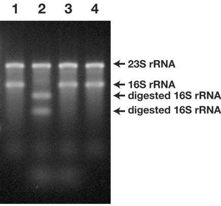
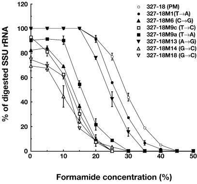
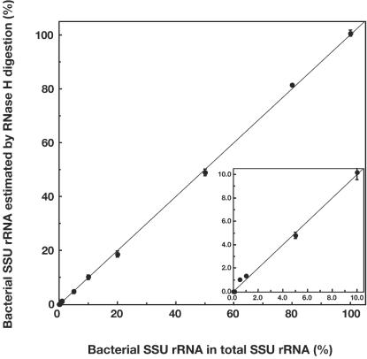
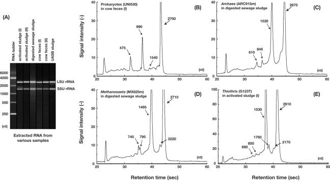
A rapid and simple approach to the small-subunit (SSU) rRNA-based quantitative detection of a specific group of microorganisms in complex ecosystems has been developed. The method employs sequence-specific cleavage of rRNA molecules with oligonucleotides and RNase H. Defined mixtures of SSU rRNAs were mixed with an oligonucleotide (referred to as a "scissor probe") that was specifically designed to hybridize with a particular site of targeted rRNA and were subsequently digested with RNase H to proceed to sequence-dependent rRNA scission at the hybridization site. Under appropriate reaction conditions, the targeted rRNAs were correctly cut into two fragments, whereas nontargeted rRNAs remained intact under the same conditions. The specificity of the cleavage could be properly adjusted by controlling the hybridization stringency between the rRNA and the oligonucleotides, i.e., by controlling either the temperature of the reaction or the formamide concentration in the hybridization-digestion buffer used for the reaction. This enabled the reliable discrimination of completely matched rRNA sequences from single-base mismatched sequences. For the detection of targeted rRNAs, the resulting RNA fragment patterns were analyzed by gel electrophoresis with nucleotide-staining fluorescent dyes in order to separate cleaved and intact rRNA molecules. The relative abundance of the targeted SSU rRNA fragments in the total SSU rRNA could easily be calculated without the use of an external standard by determining the signal intensity of individual SSU rRNA bands in the electropherogram. This approach provides a fast and easy means of identification, detection, and quantification of a particular group of microbes in clinical and environmental specimens based on rRNA.


Figures

FIG. 1.
FIG. 1.
Flow diagram showing the concept of sequence-specific digestion of SSU rRNA with oligonucleotides (scissor probes) and RNase H.
FIG. 2.
FIG. 2.
Cleavage of the 16S rRNA of E. coli with oligonucleotides (probe 530-16) and RNase H, as resolved in 1.5% agarose gel. Lanes: 1, whole E. coli RNA before digestion with RNase H; 2, digested RNA in the presence of oligonucleotides and RNase H; 3, digested RNA in hybridization-digestion buffer from which only RNase H was eliminated; 4, digested RNA in the buffer from which only the oligonucleotides had been removed. Lanes 3 and 4 demonstrate that the rRNA cleavage reaction does not occur in the absence of either the oligonucleotides or the RNase H. The digestion reactions were performed at 55°C for 15 min under the conditions described in Materials and Methods.
FIG. 3.
FIG. 3.
Effect of oligonucleotide type (G+C% and nucleotide length) on the 16S rRNA cleavage reaction. (A) Electropherogram of E. coli RNA digested with the 907-16 probe at 41°C, as resolved by an Agilent 2100 bioanalyzer with an RNA 6000 nano kit. Numbers with arrows indicate approximate estimates of the molecular weight of each peak (unit, nt). A gel-like image of the electropherogram is also shown in the graph; lane 1, RNA 6000 ladder marker (TaKaRa); lane 2, digested E. coli RNA fragments. (B) Temperature dependence of the rRNA cleavage reaction with the 907 probes. Percentages of cleaved 16S rRNA in the total 16S rRNA were directly estimated based on the peak areas of intact and cleaved 16S rRNA fragments in the electro- pherograms, and the percentages were plotted with the hybridization and digestion temperatures at which the respective reactions were performed. Error bars indicate the standard deviation of duplicate determinations. (C) Temperature dependence of the rRNA cleavage reaction with the 530 probes. Percentages of cleaved 16S rRNA in the total 16S rRNA were calculated in the same manner used for the graph in panel B and were plotted along with the hybridization and digestion temperatures used. Error bars indicate the standard deviation of duplicate determinations.
FIG. 4.
FIG. 4.
Effect of formamide concentration in hybridization-digestion buffer on rRNA scission. E. coli total RNA was cleaved with 327 scissor probes by using hybridization-digestion buffer containing different concentrations of formamide (%) (hybridization-digestion temperature, 50°C). Top panel, gel-like images of electropherograms of E. coli RNA cleaved with the 327-18 probe with different formamide concentrations. Bottom panel, formamide dependence of the rRNA cleavage reaction with the 327 probes. The percentages of cleaved 16S rRNA of the total 16S rRNA were estimated as described in the legend of Fig. 3 and were plotted together with the formamide concentrations used for creating the hybridization-digestion buffer. Error bars indicate the standard deviation of duplicate determinations.
FIG. 5.
FIG. 5.
Effect of single-base mismatches between oligonucleotides and E. coli 16S rRNA on the rRNA scission reaction. A PM probe (327-18) and MM probes containing a single-base MM at different positions and of different types were used for the cleavage of E. coli whole RNA at different formamide concentrations.
FIG. 6.
FIG. 6.
Probe dissociation curves of scissor probes under increasingly stringent hybridization and digestion conditions for the cleavage reactions. For each graph, data points indicate percentages of cleaved 16S rRNA in the total 16S rRNA estimated from electropherograms of RNA fragments with duplicate determinations (error bars indicate standard deviations). (A) Probe EUB338, specific for the domain Bacteria; (B) probe ARC915m, specific for the domain Archaea; (C) probe MX825m, specific for the genus Methanosaeta; (D) probe G123T, specific for the genus Thiothrix. In all of the experiments, in vitro-transcribed 16S rRNAs of each representative microbe were used for the digestion. For each probe, the probe sequences and the corresponding target sequences of the 16S rRNA of the tested organisms are indicated; dashes in the nontargeted rRNA sequences represent nucleotides identical to those of the targeted rRNA sequences. The vertical dotted lines indicate the optimum formamide concentrations for individual probes.
FIG. 7.
FIG. 7.
Quantitative detection of bacterial (E. coli) 16S rRNA molecules with the sequence-dependent rRNA cleavage method in artificially mixed 16S rRNA transcripts containing E. coli 16S rRNA and M. concilii 16S rRNA. Probe EUB338 was used as the scissor probe with a formamide concentration of 20% for the hybridization-digestion buffer. Defined (actual) percentages of E. coli rRNA in the total rRNA are plotted along the x axis, whereas the measured values of the percentages obtained by the present methods are shown along the y axis. The values on the y axis were estimated from the electropherograms of digested RNA, with corrections made with a cleavage coefficient of 0.96.
FIG. 8.
FIG. 8.
Application of the sequence-specific rRNA cleavage method to quantify microbes in actual community samples. (A) Gel-like images of total RNA extracted from various community samples showing virtually intact rRNA peaks, as resolved by an Agilent 2100 bioanalyzer. (B to E) Electropherograms of community RNAs digested with group-specific scissor probes and RNase H. Digestion of total RNA from cow feces (I) with the UNI530 probe specific for virtually all prokaryotes (B), from digested sewage sludge with the ARC915m probe specific for Archaea (C), from digested sewage sludge with the MX825m probe specific for the genus Methanosaeta (D), and from activated sludge (I) with the G123T probe specific for the genus Thiothrix (E) are shown. Numbers with arrows indicate approximate estimates of the molecular size (in nucleotides) of each peak.

Similar articles

Cited by

References

    1. Afonina, E. I., N. V. Chichkova, and A. A. Bogdanov. 1991. RNA-RNA and RNA-protein interactions in 30S ribosomal subunits: association of 16S rRNA fragments in the presence of ribosomal proteins. FEBS Lett. 283:251-254. - PubMed
    1. Amann, R., B. M. Fuchs, and S. Behrens. 2001. The identification of microorganisms by fluorescence in situ hybridization. Curr. Opin. Biotechnol. 12:231-236. - PubMed
    1. Amann, R., and W. Ludwig. 2000. Ribosomal RNA-targeted nucleic acid probes for studies in microbial ecology. FEMS Microbiol. Rev. 24:555-565. - PubMed
    1. Amann, R. I. 1995. In situ identification of micro-organisms by whole cell hybridization with rRNA-targeted nucleic acid probes, p. 1-15. In A. D. L. Akkermans and J. D. van Elass (ed.), Molecular microbial ecology manual. Kluwer Academic Publishers, London, United Kingdom.
    1. Amann, R. I., W. Gish, W. Miller, E. W. Myers, and D. J. Lipman. 1990. Combination of 16S rRNA-targeted oligonucleotide probes with flow cytometry for analyzing mixed microbial populations. Appl. Environ. Microbiol. 56:1919-1925. - PMC - PubMed

Publication types

LinkOut - more resources