. 2004 Jun 7;165(5):609-15.
doi: 10.1083/jcb.200403014.

Cell cycle progression after cleavage failure: mammalian somatic cells do not possess a "tetraploidy checkpoint"

Affiliations

Cell cycle progression after cleavage failure: mammalian somatic cells do not possess a "tetraploidy checkpoint"

Yumi Uetake et al. J Cell Biol. .

Abstract

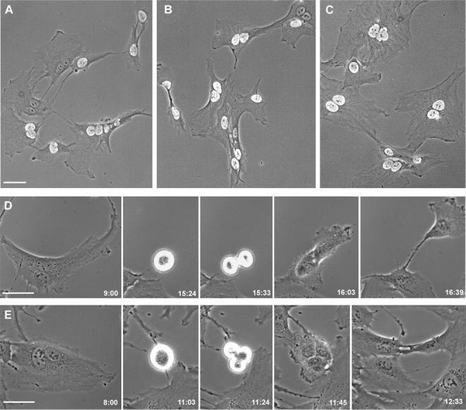
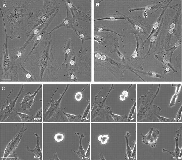
Failure of cells to cleave at the end of mitosis is dangerous to the organism because it immediately produces tetraploidy and centrosome amplification, which is thought to produce genetic imbalances. Using normal human and rat cells, we reexamined the basis for the attractive and increasingly accepted proposal that normal mammalian cells have a "tetraploidy checkpoint" that arrests binucleate cells in G1, thereby preventing their propagation. Using 10 microM cytochalasin to block cleavage, we confirm that most binucleate cells arrest in G1. However, when we use lower concentrations of cytochalasin, we find that binucleate cells undergo DNA synthesis and later proceed through mitosis in >80% of the cases for the hTERT-RPE1 human cell line, primary human fibroblasts, and the REF52 cell line. These observations provide a functional demonstration that the tetraploidy checkpoint does not exist in normal mammalian somatic cells.


Figures

Figure 1.
Figure 1.
hTERT-RPE1 cells. (A–C) Overlaid phase and fluorescence images showing BrdU incorporation in mononucleate and binucleate cells. (A) Cells previously treated with 5 μM cytochalasin D and cultured on bare glass. Some binucleates have incorporated BrdU and others have not. (B) Cells previously treated with 5 μM cytochalasin D and cultured on fibronectin-coated glass. Binucleates have incorporated BrdU. (C) Cells previously treated with 0.5 μM cytochalasin D and cultured on bare glass. Both mononucleate and binucleate cells have incorporated BrdU. (D) Cells previously treated with 0.5 μM cytochalasin D to block cleavage and cultured on bare glass (images taken from Video 2, available at http://www.jcb.org/cgi/content/full/jcb.200403014/DC1). Frames from a time-lapse video sequence showing a binucleate cell entering and progressing through mitosis. This cell divides into two despite the presence of four centrosomes. (E) Cells previously treated with blebbistatin and cultured on bare glass (images taken from Video 3). Frames from a video sequence showing a binucleate cell entering and progressing through mitosis. Phase-contrast microscopy. Times are in h:min after drug removal. Bars, 50 μm.
Figure 2.
Figure 2.
REF52 cells. (A and B) Overlaid phase and fluorescence images showing BrdU incorporation in mononucleate and binucleate cells. (A) Cells were previously treated with 0.5 μM cytochalasin D and cultured on bare glass. Mononucleate cells have incorporated BrdU, whereas the binucleates have not. (B) Cells treated with 0.5 μM cytochalasin D and cultured on fibronectin-coated glass. Both the mononucleate and binucleate cells have incorporated BrdU. (C) Cells previously treated with 0.5 μM cytochalasin D and cultured on fibronectin-coated glass (images taken from Video 4, available at http://www.jcb.org/cgi/content/full/jcb.200403014/DC1). Frames from a video sequence of two binucleate cells in the same field progressing through mitosis. The first to enter mitosis (top row) divides into two, whereas the second (bottom row) divides into three. Phase-contrast microscopy. Times are in h:min after cytochalasin D removal. Bars, 50 μm.
Figure 3.
Figure 3.
Primary human fibroblasts. (A and B) Overlaid phase and fluorescence images showing BrdU incorporation in mononucleate and binucleate cells. (A) Cells previously treated with 0.5 μM cytochalasin D and cultured on bare glass. Binucleate cells incorporate BrdU. (B) Cells previously treated with 0.5 μM cytochalasin D and cultured on fibronectin-coated glass. Binucleate cells incorporate BrdU. (C) Cells previously treated with 0.5 μM cytochalasin D and cultured on bare glass (images taken from Video 5, available at http://www.jcb.org/cgi/content/full/jcb.200403014/DC1). Frames from a video sequence of a binucleate cell progressing through mitosis. Phase-contrast microscopy. Times are in h:min after cytochalasin D removal. Bars, 50 μm.

Comment in

References

    1. Ailenberg, M., and M. Silverman. 2003. Cytochalasin D disruption of actin filaments in 3T3 cells produces an anti-apoptotic response by activating gelatinase A extracellularly and initiating intracellular survival signals. Biochim. Biophys. Acta. 1593:249–258. - PubMed
    1. Andreassen, P.R., O.D. Lohez, F.B. Lacroix, and R.L. Margolis. 2001. Tetraploid state induces p53-dependent arrest of nontransformed mammalian cells in G1. Mol. Biol. Cell. 12:1315–1328. - PMC - PubMed
    1. Bohmer, R.M., E. Scharf, and R.K. Assoian. 1996. Cytoskeletal integrity is required throughout the mitogen stimulation phase of the cell cycle and mediates the anchorage-dependent expression of cyclin D1. Mol. Biol. Cell. 7:101–111. - PMC - PubMed
    1. Borel, F., O.D. Lohez, F.B. Lacroix, and R.L. Margolis. 2002. Multiple centrosomes arise from tetraploidy checkpoint failure and mitotic centrosome clusters in p53 and RB pocket protein-compromised cells. Proc. Natl. Acad. Sci. USA. 99:9819–9824. - PMC - PubMed
    1. Brinkley, B.R. 2001. Managing the centrosome numbers game: from chaos to stability in cancer cell division. Trends Cell Biol. 11:18–21. - PubMed

Publication types