. 2004 Jun 7;165(5):723-34.
doi: 10.1083/jcb.200312172.

Integrin alphaVbeta6-mediated activation of latent TGF-beta requires the latent TGF-beta binding protein-1

Affiliations

Integrin alphaVbeta6-mediated activation of latent TGF-beta requires the latent TGF-beta binding protein-1

Justin P Annes et al. J Cell Biol. .

Abstract

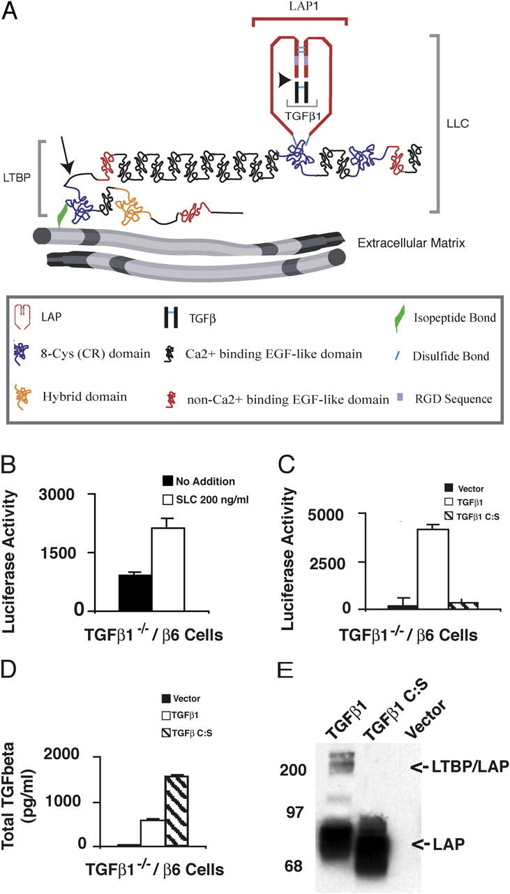
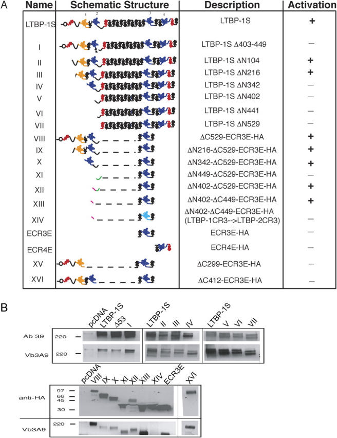
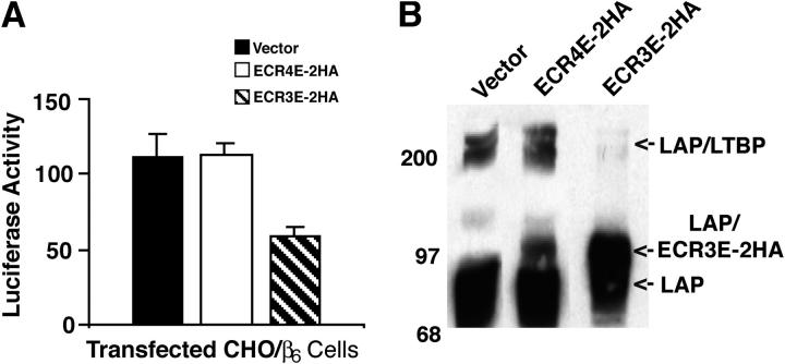
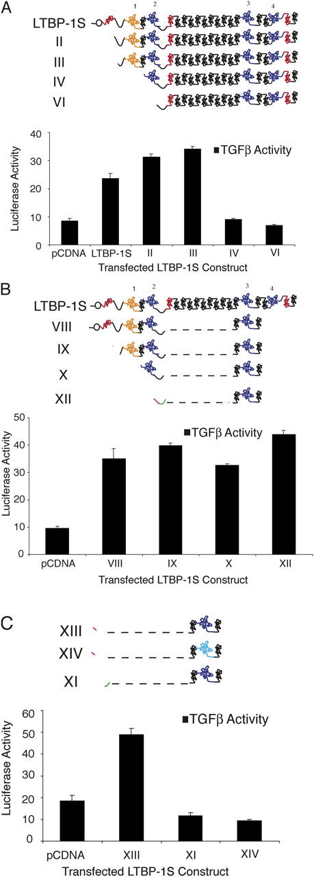
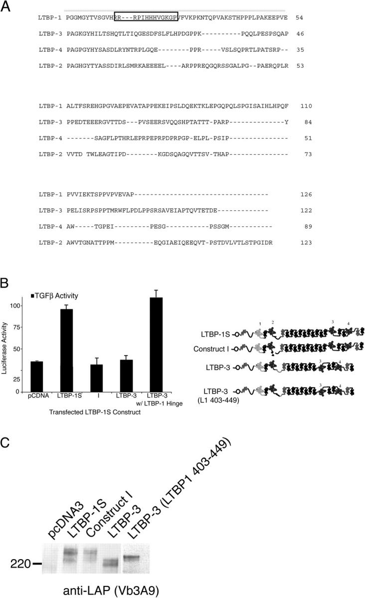
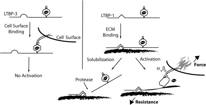
Transforming growth factor-betas (TGF-beta) are secreted as inactive complexes containing the TGF-beta, the TGF-beta propeptide, also called the latency-associated protein (LAP), and the latent TGF-beta binding protein (LTBP). Extracellular activation of this complex is a critical but incompletely understood step in TGF-beta regulation. We have investigated the role of LTBP in modulating TGF-beta generation by the integrin alphaVbeta6. We show that even though alphavbeta6 recognizes an RGD on LAP, LTBP-1 is required for alphaVbeta6-mediated latent TGF-beta activation. The domains of LTBP-1 necessary for activation include the TGF-beta propeptide-binding domain and a basic amino acid sequence (hinge domain) with ECM targeting properties. Our results demonstrate an LTBP-1 isoform-specific function in alphaVbeta6-mediated latent TGF-beta activation; LTBP-3 is unable to substitute for LTBP-1 in this assay. The results reveal a functional role for LTBP-1 in latent TGF-beta activation and suggest that activation of specific latent complexes is regulated by distinct mechanisms that may be determined by the LTBP isoform and its potential interaction with the matrix.


Figures

Figure 1.
Figure 1.
αVβ6-mediated latent TGF-β activation of SLC and TGF-β1 C:S. (A) The TGF-β large latent complex (LLC). The LLC is comprised of TGF-β (black), LAP (red) and LTBP. TGF-β and LAP are proteolytically processed at the site indicated by the arrowhead. LAP and LTBP are joined by disulfide bonds (light blue lines). The LLC is covalently linked by tTGase to the ECM via a glutamine-lysine isopeptide bond (green) near the NH2 terminus of LTBP. The hinge domain (arrow) of LTBP is a protease-sensitive region. (B) TGF-β1−/− cells that express the β-integrin subunit (TGF-β−/−6 cells) were co-cultured with TMLC that produce luciferase in response to TGF-β. Co-cultures contained either no addition or recombinant SLC (200 ng/ml). After 16–24 h, cell lysates were collected and luciferase activity measured. The errors bars represent the SEM from a single experiment performed in duplicate. This experiment was repeated multiple times with similar results. (C) TGF-β−/−6 cells were transfected with an empty vector, TGF-β1 cDNA, or TGF-β1 C:S cDNA and co-cultured with TMLCs for 16–24 h. The cell lysates were assayed for luciferase activity. (D) The transfected cells (C) were also used to generate conditioned media (CM) for 16–24 h. CM was collected, heated to 80°C for 10 min, diluted 10-fold, and added to TMLC. In separate wells, various concentrations of recombinant TGF-β were added to TMLCs to generate a TGF-β standard curve. The amount of TGF-β present in the CM was determined based upon the standard curve. Luciferase assays were performed in triplicate and the SD of a single experiment is shown. (C and D) The errors bars represent the SD of a single experiment that was performed in triplicate. These experiments were repeated multiple times with similar results. (E) The CM generated by the transfected cells was also used for Western blotting. Protein bands were revealed with Vb3A9 (anti-LAP).
Figure 2.
Figure 2.
Structure and expression of LTBP constructs. A number of LTBP-1S–derived expression constructs are used throughout this paper. (A) These constructs are schematically represented with their name, description, and ability to support latent TGF-β activation given. The highly modular structure of LTBP-1 is represented with EGF-like domains in red (noncalcium binding) and black (calcium binding), and CR domains shown in yellow (hybrid) and blue. The mutated CR3 domain of construct XIV (light blue) has been altered to resemble the non TGF-β–binding CR3 domain of LTBP-2. (B) The expression constructs shown schematically in A were cotransfected with simian proTGF-β1 into CHO cells and the conditioned media were collected and probed with Ab39 (anti–LTBP-1S), Vb3A9 (anti-LAP) and anti-HA, as appropriate, to demonstrate secretion and complex formation with LAP.
Figure 3.
Figure 3.
Affect of ECR3E on αVβ6-mediated latent TGF-β activation and LLC formation. CHO/β6 cells were transduced with empty, ECR3E- or ECR4E-expressing viruses. (A) The transduced cells were co-cultured with TGF-β-reporter TMLCs for 16–24 h before harvesting cell lysates and measuring luciferase activity. Experiments were performed in triplicate and the SDs of a single experiment are given. The errors bars represent the SD of a single experiment that was performed in triplicate. This experiment was repeated multiple times with similar results. (B) The transduced cells were transiently transfected with a TGF-β1 cDNA expression vector and allowed to generate CM for 16–24 h. The media were used for Western blotting. The reactive bands were revealed with an anti-LAP antibody (Vb3A9).
Figure 4.
Figure 4.
LTBP-1S–derived expression constructs rescue TGF-β activation. (A) CHO- β6/ECR3E cells were cotransfected with NH2-terminal deletion constructs and wild-type TGF-β1 before co-culture with TGF-β reporter TMLCs. After 16–24 h, the cell lysates were collected and luciferase activity measured. Similar experiments were conducted with additional LTBP-1S–derived expression constructs in B and C. In C, the size of the active hinge region (green) has been enlarged relative to other sequences for illustration purposes. In all cases, secretion of the transfected constructs and TGF-β complex formation was demonstrated (not depicted). Experiments were performed in triplicate. The error bars represent the SD from a single experiment.
Figure 5.
Figure 5.
Activation and LLC formation of LTBP-1S and LTBP-3. (A) Alignment of the hinge domains of LTBP-1, -2, -3, and -4 using MAP (http://searchlauncher.bcm.tmc.edu/multi-align/multi-align.html). Amino acids 403–449 of LTBP-1S are overlined. The boxed amino acid sequence identifies a putative GAG-binding sequence. (B) CHO-β6/ECR3E cells were cotransfected with either full-length TGF-β1 and LTBP-1S–derived expression constructs or LTBP-3–based expression constructs before co-culture with TGF-β reporter TMLCs. After 16–24 h, cell lysates were harvested and luciferase activity measured. The error bars represent the SD of a single experiment that was performed in triplicate. This experiment was repeated multiple times with similar results. (C) Conditioned media from the same transfected cells were collected and subjected to Western blotting using Vb3A9 to reveal reactive protein bands.
Figure 6.
Figure 6.
Activation of artificially localized latent TGF-β. (A) CHO cells that stably express β6-integrin (bars 1 and 5), β6-integrin and ECR3E (bars 2 and 6), ECR3E (bars 3 and 7), or untransfected (bars 4 and 8) were co-cultured with TMLCs on mock-coated or HA-antibody–coated wells. After 16–24 h, cell lysates were collected and assayed for luciferase activity. (B) The wells of a 96-well microtiter plate were coated with an anti-HA or control antibody, or anti-αvβ6 at various concentrations. CHO-ECR3E/β6 cells and TMLCs were co-cultured on these wells. After 16–24 h, the cell lysates were collected and the luciferase activity measured. Each experimental condition was performed in triplicate. The SD from a single experiment is shown.
Figure 7.
Figure 7.
Activation of ECM deposited latent TGF-β. CHO cells stably transfected with ECR3E or LTBP-1S (5 × 10⁴) were plated in a 96-well plate for 48 h before removal with PBS/20 mM EDTA. SW480-ECR3E/β6 or CHO-ECR3E/β6 cells and TMLCs were added to these wells. After 16–24 h, cell lysates were collected and luciferase activity measured. All experimental conditions were performed in triplicate. The SD from a single experiment is shown.
Figure 8.
Figure 8.
Schematic representation of αVβ6-mediated latent TGF-β activation. TGF-β is secreted in a complex with a variety of LTBP isoforms and splice variants. The highly variable primary sequence of the hinge domain localizes latent TGF-β in the extracellular environment. Importantly, the hinge domain of LTBP-1 functions in a capacity that is not replicated by the hinge domain of LTBP-3. Once latent TGF-β is fixed in the ECM, the integrin αVβ6 binds LAP and generates a retractile force. The magnitude of this force is related to the resistance garnered through association of the latent complex with the ECM. Once the force generated by the integrin exceeds a threshold, biologically active TGF-β is made available. Release of the latent complex from its association with the ECM, for example by proteases, is predicted to prevent αVβ6-mediated latent TGF-β activation as integrin retraction will no longer be resisted.

Similar articles

Cited by

References

    1. Abe, M., J.G. Harpel, C.N. Metz, I. Nunes, D.J. Loskutoff, and D.B. Rifkin. 1994. An assay for transforming growth factor-beta using cells transfected with a plasminogen activator inhibitor-1 promoter-luciferase construct. Anal. Biochem. 216:276–284. - PubMed
    1. Abe, M., N. Oda, and Y. Sato. 1998. Cell-associated activation of latent transforming growth factor-beta by calpain. J. Cell. Physiol. 174:186–193. - PubMed
    1. Annes, J.P., J.S. Munger, and D.B. Rifkin. 2003. Making sense of latent TGFbeta activation. J. Cell Sci. 116:217–224. - PubMed
    1. Annes, J.P., D.B. Rifkin, and J.S. Munger. 2002. The integrin alphaVbeta6 binds and activates latent TGFbeta3. FEBS Lett. 511:65–68. - PubMed
    1. Blobe, G.C., W.P. Schiemann, and H.F. Lodish. 2000. Role of transforming growth factor beta in human disease. N. Engl. J. Med. 342:1350–1358. - PubMed

Publication types

MeSH terms