. 2004 Jun 7;199(11):1467-77.
doi: 10.1084/jem.20040180.

CD25+ CD4+ T cells, expanded with dendritic cells presenting a single autoantigenic peptide, suppress autoimmune diabetes

Affiliations

CD25+ CD4+ T cells, expanded with dendritic cells presenting a single autoantigenic peptide, suppress autoimmune diabetes

Kristin V Tarbell et al. J Exp Med. .

Abstract

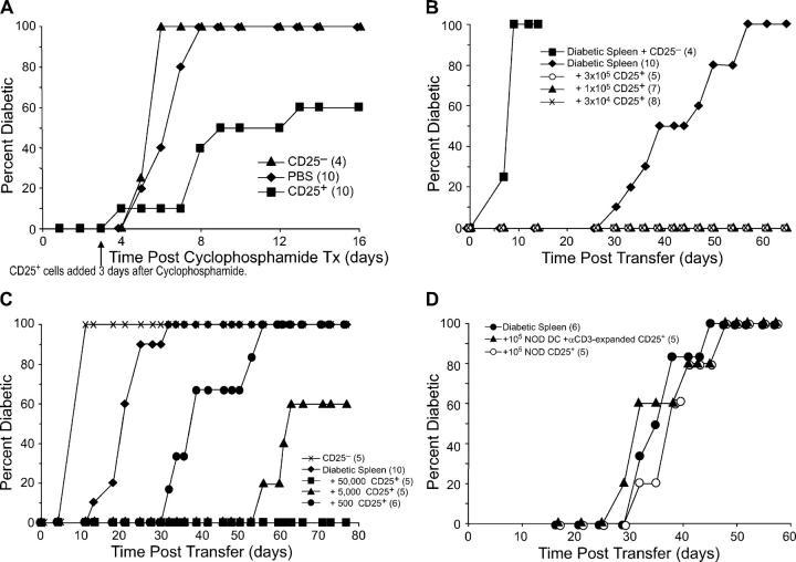
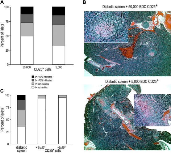
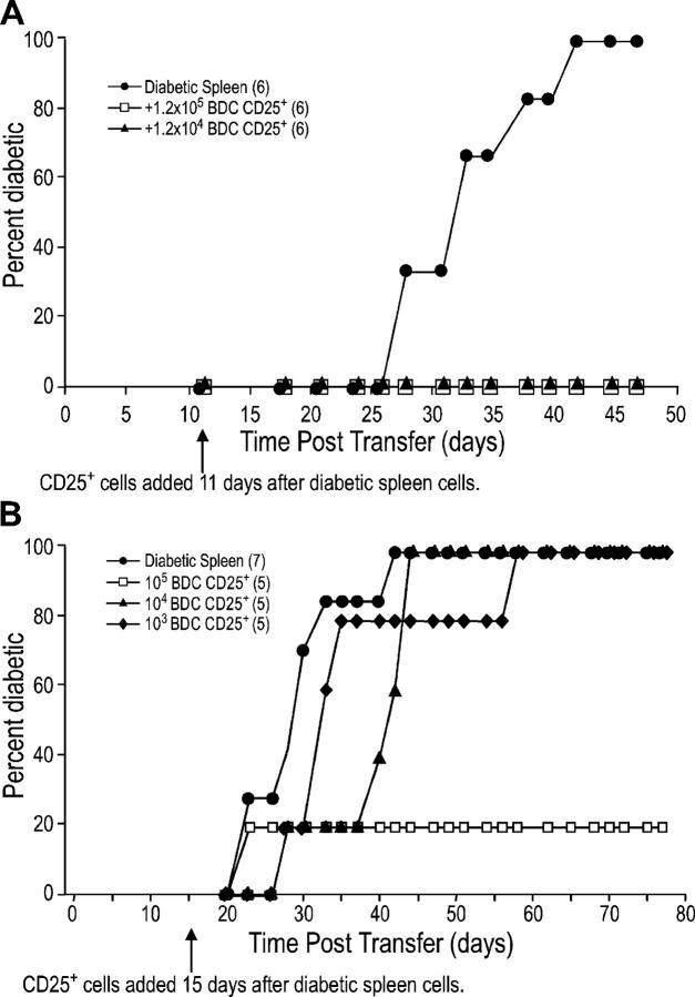
In the nonobese diabetic (NOD) mouse model of type 1 diabetes, the immune system recognizes many autoantigens expressed in pancreatic islet beta cells. To silence autoimmunity, we used dendritic cells (DCs) from NOD mice to expand CD25+ CD4+ suppressor T cells from BDC2.5 mice, which are specific for a single islet autoantigen. The expanded T cells were more suppressive in vitro than their freshly isolated counterparts, indicating that DCs from autoimmune mice can increase the number and function of antigen-specific, CD25+ CD4+ regulatory T cells. Importantly, only 5,000 expanded CD25+ CD4+ BDC2.5 T cells could block autoimmunity caused by diabetogenic T cells in NOD mice, whereas 10(5) polyclonal, CD25+ CD4+ T cells from NOD mice were inactive. When islets were examined in treated mice, insulitis development was blocked at early (3 wk) but not later (11 wk) time points. The expanded CD25+ CD4+ BDC2.5 T cells were effective even if administered 14 d after the diabetogenic T cells. Our data indicate that DCs can generate CD25+ CD4+ T cells that suppress autoimmune disease in vivo. This might be harnessed as a new avenue for immunotherapy, especially because CD25+ CD4+ regulatory cells responsive to a single autoantigen can inhibit diabetes mediated by reactivity to multiple antigens.


Figures

Figure 1.
Figure 1.
NOD DCs induce growth of CD25+ CD4+ T cells from NOD.BDC2.5 or NOD mice. (A) In vitro–derived NOD DCs were stained with antibodies specific for CD86 and MHC class II before (left) and after (right) magnetic bead enrichment of CD86+ cells. (B) CD25+ CD4+ or CD25 CD4+ T cells sorted from BDC2.5 TCR transgenic mice were cultured with CD86+ NOD DCs with and without 30 ng ml−1 BDC peptide and IL-2. In the same experiment, NOD spleen cells were used with IL-2, with and without BDC peptide (right). A 12-h [3H]thymidine pulse was given on day 3. (C) Same as B, but the dose of BDC peptide was 100 ng ml−1 and the fold increase in T cell numbers was monitored by counting on days 3, 5, and 7. (D) CD25+ or CD25 CD4+ T cells were isolated from NOD mice and cultured with NOD CD86+ DCs, with and without anti-CD3 and IL-2 as indicated. Proliferation was determined by [3H]thymidine incorporation on day 3. (E) As in D, but cells were counted on days 3, 5, and 7, and the fold increase in cell numbers was calculated. One result of at least three similar experiments is shown.
Figure 2.
Figure 2.
Phenotype of DC-expanded T cells. Expression of BDC2.5 clonotype (left), TCR Vα2 (middle), and CD62L (right) on CD25+ or CD25 CD4+ T cells cultured under the conditions indicated. For the BDC2.5 clonotype, the isotype control peak is shown by the dashed line. For Vα2 and CD62L, the percentage of cells in the indicated gates is shown. One result of at least three similar experiments is shown.
Figure 3.
Figure 3.
BDC2.5 CD25+ CD4+ T cells proliferate in vivo. CFSE-labeled BDC2.5 CD25 CD4+ (left) or CD25+ CD4+ (right) T cells were injected into NOD mice. 1 d later, either DCs without antigen (top) or BDC peptide–pulsed DCs (middle and bottom) were injected s.c. 3 d after antigen delivery, the injected >1,000 CFSE-labeled clonotype+ cells from draining (top and middle) or distal (bottom) lymph nodes were assessed for proliferation by flow cytometry, gating on CD4+ lymphocytes. The percentages in the dividing and nondivding populations of clonotype+ cells is shown.
Figure 4.
Figure 4.
DC-expanded CD25+ CD4+ T cells suppress proliferation better than unexpanded CD25+ CD4+ T cells. (A) CD25+ CD4+ T cells from NOD.BDC2.5 mice were expanded for 7 d with irradiated NOD DCs and BDC peptide and IL-2 as indicated. 104 freshly isolated, sorted CD25 CD4+ T cells from BDC2.5 mice were cultured with NOD spleen cells, 30 ng/ml BDC peptide, and either freshly sorted CD25+ CD4+ or the indicated DC-expanded CD25+ CD4+ populations, at the ratios indicated. After 72 h, proliferation was assessed by [3H]thymidine incorporation during a 12-h pulse. One representative result from at least three is shown. (B) Same as A, but both CD25+ and CD25 CD4+ T cells were isolated from NOD mice, and anti-CD3 was used as TCR stimulus instead of BDC peptide in both expansion and suppression cultures. One representative result from at least three is shown.
Figure 5.
Figure 5.
Expanded CD25+ CD4+ T cells function in vivo to suppress development of diabetes. (A) 4–6-wk-old NOD.BDC2.5 mice were given cyclophosphamide i.p. 3 d later, either 5 × 10⁵ DC-expanded CD25+ CD4+ T cells or CD25 CD4+ cells were injected i.v. (B) NOD.scid females were injected with 3 × 10⁶ spleen cells from a diabetic NOD female and either nothing or the indicated numbers of DC-expanded CD25+ CD4+ T cells or 3 × 10⁵ CD25 CD4+ cells from BDC2.5 mice. (C) NOD.scid females were injected with either 4 × 10⁵ CD25 CD4+ cells from BDC2.5 mice, or 8 × 10⁶ spleen cells from a diabetic NOD female and either nothing or the indicated numbers of DC-expanded CD25+ CD4+ T cells from BDC2.5 mice. The difference between diabetic spleen alone to diabetic spleen plus 500 CD25+ CD4+ cells was significant (P = 0.002), as was diabetic spleen to diabetic spleen plus 5,000 CD25+ CD4+ cells (P = 0.002). One representative result from two experiments is shown. (D) NOD.scid females were injected with 8 × 10⁶ diabetic spleen cells alone or with 105 freshly isolated or DC/αCD3-expanded CD25+ CD4+ T cells from NOD mice. The number of mice in each group is indicated in parentheses.
Figure 6.
Figure 6.
Protected mice have lymphocytic infiltrates in the pancreas. (A) Pancreata from mice that did not develop diabetes by day 80 after transfer in the experiment shown in Fig. 5 C were scored for insulitis. 150 islets from 5 mice were scored from the group that received 50,000 DC-expanded BDC CD25+ CD4+ T cells, and 48 islets from 2 mice were scored from the group that received 5,000 cells. (B) Representative fields for a mouse from the group that received 5,000 (top) or 50,000 (bottom) suppressor T cells. Large field is 5×; inset is 20×. (C) In a separate experiment, pancreata from mice 28 d after transfer were scored for insulitis. At least 50 islets from 2–3 mice were scored from each group.
Figure 7.
Figure 7.
BDC2.5 CD25+ CD4+ T cells can still regulate diabetes when given after diabetogenic cells. (A) NOD.scid females were injected with 8 × 10⁶ diabetic spleen cells and 11 d later were injected with either PBS or the indicated number of DC-expanded CD25+ CD4+ T cells from BDC2.5 mice. The difference between diabetic spleen alone to diabetic spleen plus 105 or 104 DC-expanded CD25+ CD4+ cells was significant (P = 0.002). (B) As in A, except 107 diabetic spleen cells were added 15 d before the indicated number of DC-expanded CD25+ CD4+ T cells from BDC2.5 mice. The p-value for diabetic spleen alone versus adding 105 BDC2.5 regulatory T cells is 0.055 and versus adding 104 BDC2.5 regulatory T cells is 0.0595. The number of mice in each group is indicated in parentheses.

References

    1. Tisch, R., and H. McDevitt. 1996. Insulin-dependent diabetes mellitus. Cell. 85:291–297. - PubMed
    1. Wong, F.S., and C.A. Janeway, Jr. 1999. Insulin-dependent diabetes mellitus and its animal models. Curr. Opin. Immunol. 11:643–647. - PubMed
    1. Makino, S., K. Kunimoto, Y. Muraoka, Y. Mizushima, K. Katagiri, and Y. Tochino. 1980. Breeding of a non-obese, diabetic strain of mice. Jikken Dobutsu. 29:1–13. - PubMed
    1. Tisch, R., X.-Y. Yang, S.M. Singer, R.S. Libiau, L. Fugger, and H.O. McDevitt. 1993. Immune response to glutamic acid decarboxylase correlates with insulitis in non-obese diabetic mice. Nature. 366:72–75. - PubMed
    1. Daniel, D., R.G. Gill, N. Schloot, and D. Wegmann. 1995. Epitope specificity, cytokine production profile and diabetogenic activity of insulin-specific T cell clones isolated from NOD mice. Eur. J. Immunol. 25:1056–1062. - PubMed

Publication types