. 2004 Jun 15;101(24):8930-5.
doi: 10.1073/pnas.0403002101. Epub 2004 Jun 7.

Myosin subfragment 1 structures reveal a partially bound nucleotide and a complex salt bridge that helps couple nucleotide and actin binding

Affiliations

Myosin subfragment 1 structures reveal a partially bound nucleotide and a complex salt bridge that helps couple nucleotide and actin binding

Dipesh Risal et al. Proc Natl Acad Sci U S A. .

Abstract

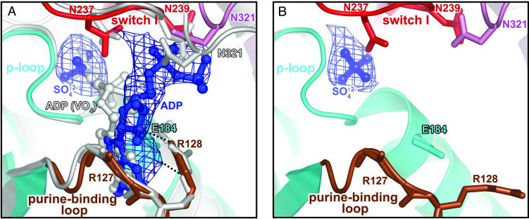
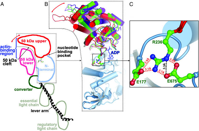
Structural studies of myosin have indicated some of the conformational changes that occur in this protein during the contractile cycle, and we have now observed a conformational change in a bound nucleotide as well. The 3.1-A x-ray structure of the scallop myosin head domain (subfragment 1) in the ADP-bound near-rigor state (lever arm =45 degrees to the helical actin axis) shows the diphosphate moiety positioned on the surface of the nucleotide-binding pocket, rather than deep within it as had been observed previously. This conformation strongly suggests a specific mode of entry and exit of the nucleotide from the nucleotide-binding pocket through the so-called "front door." In addition, using a variety of scallop structures, including a relatively high-resolution 2.75-A nucleotide-free near-rigor structure, we have identified a conserved complex salt bridge connecting the 50-kDa upper and N-terminal subdomains. This salt bridge is present only in crystal structures of muscle myosin isoforms that exhibit a strong reciprocal relationship (also known as coupling) between actin and nucleotide affinity.


Figures

Fig. 1.
Fig. 1.
Overview of the nucleotide-binding pocket. (A) ADP (blue) in the scallop myosin near-rigor structure adopts a partially bound conformation at the front door of the nucleotide-binding pocket. For comparison, the fully bound ADP from the S1–ADP·VO4 complex in the scallop prepower-stroke structure (PDB ID code 1QVI) is shown in light gray. Also shown is an FoFc simulated-annealing electron density map at a 1.0 σ contour level, calculated after omitting ADP and sulfate. Residues in switch I (red) and N321 (pink) from the 50-kDa upper subdomain that are involved in weak interactions (see text) with the ADP diphosphate moiety are shown in stick representation. The same residues from the prepower-stroke conformation structure are shown in light gray. The only major difference between the two structures in the nucleotide-binding pocket of the enzyme is that N321 (50-kDa upper subdomain) makes closer contact with the bound nucleotide in the prepower-stroke structure. The side chain of R127 and the hydrogen bond between its main-chain amide and the ADP ribose O2′ are not shown for clarity. The P-loop and purine-binding loop are shown in cyan and brown, respectively. E184 (cyan sticks) and R128 (brown sticks) in the two loops form a salt bridge (dashed lines) that protects one face of the ADP base from solvent. (B) Corresponding view of the ScS1–SO4 structure, with an FoFc simulated-annealing electron density map at a 2.0 σ contour level, calculated after omitting the sulfate. The E184/R128 salt bridge is not formed in the absence of a nucleotide, supporting the role of R128 in nucleotide recruitment.
Fig. 2.
Fig. 2.
Rotations of the 50-kDa upper subdomain in the three conformational states of scallop myosin. The 50-kDa upper subdomain rotates as a rigid body with respect to the N-terminal subdomain, with the center of rotation near R236 of switch I. (A) Schematic representation of the different subdomains in the ScS1–ADP structure. Red arrow indicates rotation of the 50-kDa upper subdomain, which closes the 50-kDa cleft when myosin binds strongly to actin (5, 6). (B) A superposition of the N-terminal subdomains (light blue) shows the relative positions of the 50-kDa upper subdomains in the present ScS1–ADP near-rigor state (red), the internally uncoupled state (green), and the prepower-stroke state (purple) structures. A red arrow indicates the rotation of the 50-kDa upper subdomain in the plane of the paper. The 50-kDa lower subdomain, the converter, and the lever arm have been omitted for clarity. ADP from the ScS1–ADP structure is shown in a blue ball-and-stick representation. Residues involved in the complex salt bridge (E177, R236, and E675) between the 50-kDa upper and N-terminal subdomains are also shown in a ball-and-stick representation. (C) Close-up view of the complex salt bridge, which is well situated to stabilize the rotation of the 50-kDa upper subdomain. All but one of the salt links have favorable geometry to form hydrogen bonds, and these are shown as red dashed lines. The additional nonhydrogen-bonded salt link is shown as a black dashed line.

References

    1. Houdusse, A., Szent-Györgyi, A. G. & Cohen, C. (2000) Proc. Natl. Acad. Sci. USA 97, 11238–11243. - PMC - PubMed
    1. Houdusse, A., Kalabokis, V. N., Himmel, D., Szent-Györgyi, A. G. & Cohen, C. (1999) Cell 97, 459–470. - PubMed
    1. Himmel, D. M., Gourinath, S., Reshetnikova, L., Shen, Y., Szent-Györgyi, A. G. & Cohen, C. (2002) Proc. Natl. Acad. Sci. USA 99, 12645–12650. - PMC - PubMed
    1. Rayment, I., Holden, H. M., Whittaker, M., Yohn, C. B., Lorenz, M., Holmes, K. C. & Milligan, R. A. (1993) Science 261, 58–65. - PubMed
    1. Volkmann, N., Hanein, D., Ouyang, G., Trybus, K. M., DeRosier, D. J. & Lowey, S. (2000) Nat. Struct. Biol. 12, 1147–1155. - PubMed

Publication types

LinkOut - more resources