

Tumor-specific Ab-mediated targeting of MHC-peptide complexes induces regression of human tumor xenografts in vivo
- PMID: 15184663
- PMCID: PMC428471
- DOI: 10.1073/pnas.0403222101
Tumor-specific Ab-mediated targeting of MHC-peptide complexes induces regression of human tumor xenografts in vivo
Abstract
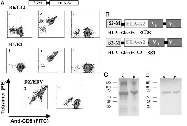
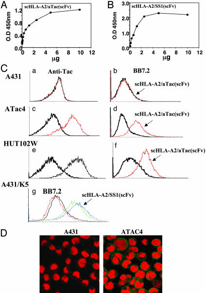
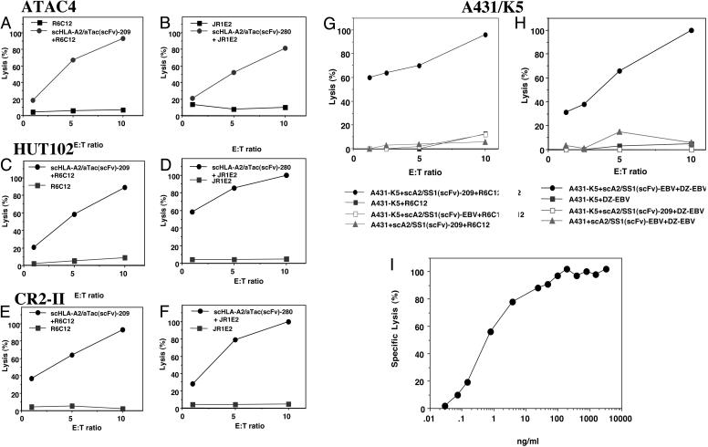
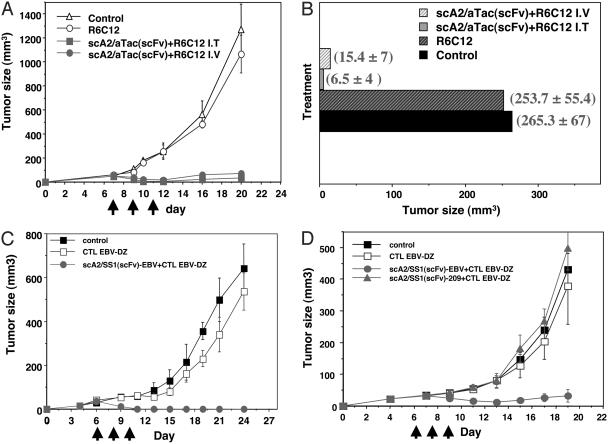
A cancer immunotherapy strategy is described herein that combines the advantage of the well established tumor targeting capabilities of high-affinity recombinant fragments of Abs with the known efficient, specific, and potent killing ability of CD8 T lymphocytes directed against highly antigenic MHC-peptide complexes. Structurally, it consists of a previously uncharacterized class of recombinant chimerical molecules created by the genetic fusion of single-chain (sc) Fv Ab fragments, specific for tumor cell surface antigens, to monomeric scHLA-A2 complexes containing immunodominant tumor- or viral-specific peptides. The fusion protein can induce very efficiently tumor cell lysis, regardless of the expression of self peptide-MHC complexes. Moreover, these molecules exhibited very potent antitumor activity in vivo in nude mice bearing preestablished human tumor xenografts. These in vitro and in vivo results suggest that recombinant scFv-MHC-peptide fusion molecules could represent an approach to immunotherapy, bridging Ab and T lymphocyte attack on cancer cells.
Figures
Similar articles
-
Selective antibody-mediated targeting of class I MHC to EGFR-expressing tumor cells induces potent antitumor CTL activity in vitro and in vivo.Int J Cancer. 2007 Jan 15;120(2):329-36. doi: 10.1002/ijc.22168. Int J Cancer. 2007. PMID: 17066453
-
Recruitment of CTL activity by tumor-specific antibody-mediated targeting of single-chain class I MHC-peptide complexes.J Immunol. 2002 Sep 15;169(6):2988-96. doi: 10.4049/jimmunol.169.6.2988. J Immunol. 2002. PMID: 12218113
-
Redirecting anti-viral CTL against cancer cells by surface targeting of monomeric MHC class I-viral peptide conjugated to antibody fragments.Cancer Immun. 2001 Mar 30;1:2. Cancer Immun. 2001. PMID: 12747763
-
CD7-specific single chain Fv immunotoxins. Design and expression.Methods Mol Biol. 2001;166:17-29. doi: 10.1385/1-59259-114-0:17. Methods Mol Biol. 2001. PMID: 11217366 Review. No abstract available.
-
DNA fusion vaccines enter the clinic.Cancer Immunol Immunother. 2011 Aug;60(8):1147-51. doi: 10.1007/s00262-011-1042-2. Epub 2011 Jun 5. Cancer Immunol Immunother. 2011. PMID: 21644035 Free PMC article. Review.
Cited by
-
Stable, soluble, high-affinity, engineered T cell receptors: novel antibody-like proteins for specific targeting of peptide antigens.Clin Exp Immunol. 2005 Dec;142(3):454-60. doi: 10.1111/j.1365-2249.2005.02929.x. Clin Exp Immunol. 2005. PMID: 16297157 Free PMC article. Review.
-
Introduction to monoclonal antibodies.Cancer Immun. 2012;12:11. Epub 2012 May 1. Cancer Immun. 2012. PMID: 22896756 Free PMC article. Review. No abstract available.
-
Antigen-specific T cell Redirectors: a nanoparticle based approach for redirecting T cells.Oncotarget. 2016 Oct 18;7(42):68503-68512. doi: 10.18632/oncotarget.11785. Oncotarget. 2016. PMID: 27602488 Free PMC article.
-
Targeted coating with antigenic peptide renders tumor cells susceptible to CD8(+) T cell-mediated killing.Mol Ther. 2013 Mar;21(3):542-53. doi: 10.1038/mt.2012.233. Epub 2012 Nov 27. Mol Ther. 2013. PMID: 23183537 Free PMC article.
-
Immunotherapies: Exploiting the Immune System for Cancer Treatment.J Immunol Res. 2018 Mar 14;2018:9585614. doi: 10.1155/2018/9585614. eCollection 2018. J Immunol Res. 2018. PMID: 29725606 Free PMC article. Review.
References
-
- McLaughlin, P., Grillo-Lopez, A. J., Link, B. K., Levy, R., Czuczman, M. S., Williams, M. E., Heyman, M. R., Bence-Bruckler, I., White, C. A., Cabanillas, F., et al. (1998) J. Clin. Oncol. 16, 2825-2833. - PubMed
-
- Cobleigh, M. A., Vogel, C. L., Tripathy, D., Robert, N. J., Scholl, S., Fehrenbacher, L., Wolter, J. M., Paton, V., Shak, S., Lieberman, G. & Slamon, D. J. (1999) J. Clin. Oncol. 17, 2639-2648. - PubMed
-
- Ferrara, N. (2002) Semin. Oncol. 29, 10-14. - PubMed
-
- Pastan, I. (1997) Biochim. Biophys. Acta 1333, C1-C6. - PubMed
-
- Lode, H. N. & Reisfeld, R. A. (2000) Immunol. Res. 21, 279-288. - PubMed
Publication types
MeSH terms
Substances
LinkOut - more resources
Full Text Sources
Other Literature Sources
Research Materials
