. 2004 Jun 15;101(24):9097-102.
doi: 10.1073/pnas.0400438101. Epub 2004 Jun 7.

Functional organization of sensory input to the olfactory bulb glomerulus analyzed by two-photon calcium imaging

Affiliations

Functional organization of sensory input to the olfactory bulb glomerulus analyzed by two-photon calcium imaging

Matt Wachowiak et al. Proc Natl Acad Sci U S A. .

Abstract

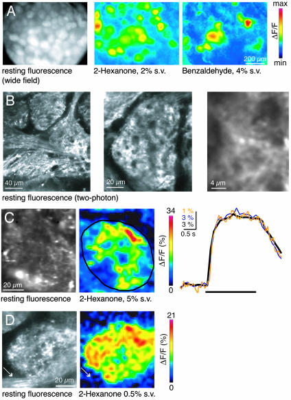
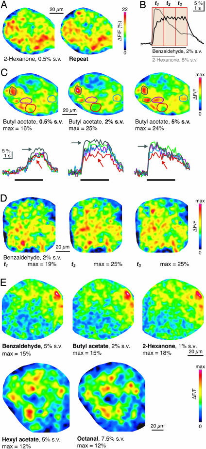
Glomeruli in the olfactory bulb are anatomically discrete modules receiving input from idiotypic olfactory sensory neurons. To examine the functional organization of sensory inputs to individual glomeruli, we loaded olfactory sensory neurons with a Ca(2+) indicator and measured odorant-evoked presynaptic Ca(2+) signals within single glomeruli by using two-photon microscopy in anaesthetized mice. Odorants evoked patterns of discrete Ca(2+) signals throughout the neuropil of a glomerulus. Across glomeruli, Ca(2+) signals occurred with equal probability in all glomerular regions. Within single glomeruli, the pattern of intraglomerular Ca(2+) signals was indistinguishable for stimuli of different duration, identity, and concentration. Moreover, the response time course of the signals was similar throughout the glomerulus. Hence, sensory inputs to individual glomeruli are spatially heterogeneous but seem to be functionally indiscriminate. These results support the view of olfactory glomeruli as functional units in representing sensory information.


Figures

Fig. 1.
Fig. 1.
Two-photon imaging reveals patterns of intraglomerular presynaptic Ca2+ signals. (A) Wide-field fluorescence imaging of glomeruli after loading of OSN axons with calcium green-1 dextran (Left) and activity patterns evoked by two odorants (Center and Right). Imaging was performed in vivo through the thinned skull in a freely breathing animal. Color scale: -1.75 to 7% for 2-hexanone, and -1 to 4% for benzaldehyde. (B) In vivo two-photon scans of different glomeruli at increasing magnification after loading of OSN axons with calcium green-1 dextran or Oregon green BAPTA-1 dextran. (C) Two-photon imaging of resting fluorescence and intraglomerular Ca2+ signals in an optical section through an individual glomerulus. (Right) Response time course in the outlined region. Yellow, wide-field imaging, single trial; blue, two-photon imaging, single trial; black, two-photon imaging, average of eight trials. (D) Odorant-evoked Ca2+ signals in an optical section through a glomerulus and innervating axon bundle (arrow).
Fig. 2.
Fig. 2.
Distribution of odorant-evoked intraglomerular Ca2+ signals. (A) High spatial resolution image of fluorescence within the glomerular neuropil (Left) and map of odorant-evoked Ca2+ signal in the same region (Right). (B) Overlay of resting fluorescence and odorant-evoked Ca2+ signal map in A, rescaled and thresholded at 40% of their maxima. (C) Temporal structure of Ca2+ signals. Resting fluorescence (Left) and odorant-evoked Ca2+ signals (Center) within a glomerulus (high magnification). The area approximated by the gray rectangle was subsequently scanned at high temporal resolution (frame rate, 62.5 Hz). (Right) Time courses were smoothed by a running average over three adjacent data points. Arrows depict uncorrelated signal fluctuations. (D) Maps of Ca2+ signals evoked by 2-hexanone (5%) at different depths in the same glomerulus. Z = 0 μm corresponds to a level just below the olfactory nerve layer. White mask outlines glomerular border. (E) Normalized average intraglomerular Ca2+ signal as a function of radial position in the glomerular cross section.
Fig. 3.
Fig. 3.
Stimulus dependence of odorant-evoked maps of intraglomerular Ca2+ signals. (A) Intraglomerular patterns of Ca2+ signals evoked by repeated stimulation with the same odorant. White mask outlines glomerular border. (B) Time course of the response of the glomerulus in D, averaged over the entire cross section. Red boxes depict the time windows used to construct the activity maps in D. Gray trace shows the response to a different odorant (2-hexanone, 5%), which evoked a different temporal response. (C) Intraglomerular activity maps evoked by increasing concentrations of the same odorant. Traces show time courses of the Ca2+ signal in the regions outlined by corresponding line colors. (D) Maps evoked by benzaldehyde (2%) in subsequent 1-s time windows during a 3-s odorant presentation. The response time course is shown in B. (E) Intraglomerular activity maps evoked by three different odorants in one glomerulus (Upper) and two different odorants in another glomerulus (Lower).
Fig. 4.
Fig. 4.
Statistical analysis of similarity between intraglomerular activity maps. (A) Average correlation (±SD) between activity maps evoked by repeated application of the same odorant in the same time window (Same), the same odorant in subsequent 1-s time windows (Diff. time), the same odorant at different concentrations (Diff. conc.), or different odorants (Diff. odor). (B) Average correlation between activity maps, after setting ΔF/F values lower than a 30% of the maximum ΔF/F in at least one of the maps in a comparison to 0.

References

    1. Mombaerts, P. (1999) Science 286, 707-711. - PubMed
    1. Hálasz, N. & Greer, C. A. (1993) J. Comp. Neurol. 337, 307-316. - PubMed
    1. Shepherd, G. M. & Greer, C. A. (1998) in The Synaptic Organization of the Brain, ed. Shepherd, G. M. (Oxford Univ. Press, New York), pp. 159-203.
    1. Le Gros Clark, W. (1957) Proc. R. Soc. London Ser. B 146, 299-319. - PubMed
    1. Kauer, J. S. & Cinelli, A. R. (1993) Microsc. Res. Tech. 24, 157-167. - PubMed

Publication types

LinkOut - more resources