Review
. 2004 May;5(5):464-9.
doi: 10.1038/sj.embor.7400146.

Structures of protein domains that create or recognize histone modifications

Affiliations
Review

Structures of protein domains that create or recognize histone modifications

Matthew J Bottomley. EMBO Rep. 2004 May.

Abstract

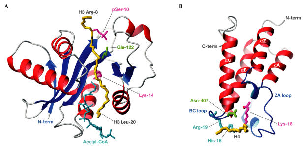
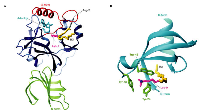
DNA is packed together with histone proteins in cell nuclei to form a compact structure called chromatin. Chromatin represents a scaffold for many genetic events and shows varying degrees of condensation, including a relatively open form (euchromatin) and a highly condensed form (heterochromatin). Enzymes such as histone acetyltransferases (HATs) and methylases covalently label the amino-termini of histones, thereby creating a 'histone code' of modifications that is interpreted by the recruitment of other proteins through recognition domains. Ultimately, this network of interacting proteins is thought to control the degree of chromatin condensation so that DNA is available when it is required for genomic processes. Reviewed here are the structures of HAT and SET domains, which mediate the acetylation and methylation of histones, respectively, and bromodomains and chromodomains, which recognize the modified histones. How these structures have increased our understanding of DNA regulation is also discussed.


Figures

Figure 1
Figure 1
The wide range of histone modifications. (A) Chromatin is formed by nucleosome subunits comprising an octameric core of histones around which ∼1.8 superhelical turns of DNA are wrapped. DNA binds to the positively charged histone surfaces through H-bonds and electrostatic interactions. In the 11-nm chromatin fibre, successive nucleosomes are separated by 10–80 base pairs of linker DNA. Histone H1, which binds to nucleosomes and adjacent linker DNA, can mediate further condensation into the 30-nm chromatin fibre. (B) The amino-termini of histones H3 and H4 protrude from the nucleosome core and contain dense clusters of modifiable residues. Residues are coloured on well-documented modification sites, with red indicating where acetylation or phosphorylation increase acidity and blue indicating methylation. Note that H3 Lys 9 (pink) can be either acetylated or methylated. Pale green shading highlights putative 'modification cassettes' and boxes indicate potential 'methyl–phos binary switches'. Further possible modifications are listed elsewhere (Felsenfeld & Groudine, 2003; Fischle et al, 2003a). (C) Histone modifications create new chemical environments. For example, lysine acetylation (red) neutralizes the positive charge of the Nζ group and introduces the carbonyl oxygen, which is a potential H-bond acceptor. Lysine methylation (blue) increases both hydrophobicity and the cationic nature of the Nζ group. Depending on the modifying enzyme, lysines can be mono-, di- or tri-methylated.
Figure 2
Figure 2
Acetyl-Lys modification and recognition. (A) The tGCN5 HAT domain with bound acetyl-CoA (cyan) and an H3 peptide spanning Arg 8 to Leu 20 (backbone in gold; Protein Data Bank (PDB) code ). Also shown are side chains of the catalytic Glu 122 (green) and H3 pSer 10, and target Lys 14 (magenta). (B) The GCN5 bromodomain bundle (red), bound to an H4 peptide spanning Ala 15 to Arg 19 (backbone in gold; PDB code ). The acetyl-Lys 16 (magenta) of H4 reaches far into the pocket that is formed by the bromodomain loops ZA and BC (blue). An H-bond forms between the acetyl oxygen atom (red) of Lys 16 and Asn 407 (green). The H4 residues His 18 and Arg 19 (cyan), in positions acetyl-Lys +2 and +3, make interactions that are specific for GCN5.
Figure 3
Figure 3
Methyl-Lys modification and recognition. (A) The SET7/9 domain complexed with AdoHcy (cyan) and an H3 peptide spanning Arg 2 to Lys 9 (backbone in gold; Protein Data Bank (PDB) code ). The H3 methyl-Lys 4 side chain (magenta) projects from the front face, through the lysine-access channel, to reach AdoHcy at the back. The SET domain (blue) contains the conserved SET-N and SET-C domains, each of three or four β-strands, a short helix and several loops, plus the variable SET-I region. The N-terminal SET7/9 domain (green) makes hydrophobic interactions with the SET domain. The SET-C flanking region (red) packs against the SET domain to form the lysine-access channel, the histone-binding groove that selects H3 through interactions with Arg 2 (grey) and hydrophobic interactions with AdoHcy, which collectively explain its importance for SET activity. (B) The HP1β chromodomain (cyan) complexed with an H3 peptide (Gln 5 to Ser 10; backbone in gold) sandwiched between strands β4 and the histone-induced strand β1 (PDB code ). The H3 di-methyl-Lys 9 side chain (magenta) is surrounded along its length by hydrophobic residues that are conserved in many chromodomains. The Lys-Nζ atom (blue) sits centrally in the three-walled aromatic cage that is formed by Tyr 24, Trp 45 and Tyr 48 (green), which can accommodate di- or tri-methyl-Lys.
None

Similar articles

Cited by

References

    1. Akhtar A, Zink D, Becker PB (2000) Chromodomains are protein–RNA interaction modules. Nature 407: 405–409 - PubMed
    1. Allfrey VG, Faulkner R, Mirsky AE (1964) Acetylation and methylation of histones and their possible role in the regulation of RNA synthesis. Proc Natl Acad Sci USA 51: 786–794 - PMC - PubMed
    1. Bauer UM, Daujat S, Nielsen SJ, Nightingale K, Kouzarides T (2002) Methylation at arginine 17 of histone H3 is linked to gene activation. EMBO Rep 3: 39–44 - PMC - PubMed
    1. Bottomley MJ, Collard MW, Huggenvik JI, Liu Z, Gibson TJ, Sattler M (2001) The SAND domain structure defines a novel DNA-binding fold in transcriptional regulation. Nat Struct Biol 8: 626–633 - PubMed
    1. Bouazoune K, Mitterweger A, Langst G, Imhof A, Akhtar A, Becker PB, Brehm A (2002) The dMi-2 chromodomains are DNA binding modules important for ATP-dependent nucleosome mobilization. EMBO J 21: 2430–2440 - PMC - PubMed

Publication types