. 2004 Dec;53(12):1146-52.
doi: 10.1007/s00262-004-0556-2.

Evidence for immune defects in breast and lung cancer patients

Affiliations

Evidence for immune defects in breast and lung cancer patients

Iuliana Caras et al. Cancer Immunol Immunother. 2004 Dec.

Abstract

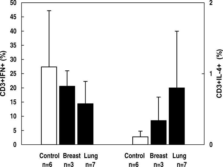
Immunosuppression is often identified in cancer patients. The aim of this study was to evaluate several immune parameters for patients with breast and lung cancer. Immunophenotyping analysis showed that the cancer patients investigated had significantly lower absolute numbers of peripheral blood lymphocytes than controls. The immunosuppression was more evident for the breast cancer subgroup. The most severe immune defect noticed was the marked impairment of IFN-gamma secretion. A shift toward the Th2 phenotype as revealed by assessment of intracellular level of IFN-gamma and IL-4 was also noticed. The secretion of proinflammatory cytokines IL-1beta and TNF-alpha in whole blood cultures was not impaired. Although the proportion of activated cells was slightly lower than in the control group, our results showed that both peripheral T lymphocytes and NK cells of cancer patients could be induced to express early activation marker CD69 after ex vivo mitogen stimulation. In conclusion, our study revealed several immune defects in cancer patients. This suggests that an appropriate immunotherapeutical approach might be used to restore compromised immune functions with beneficial effects on both antitumor and general immunity.


Figures

Fig. 1
Fig. 1
Proportion of IFN-γ+ and IL-4+ T lymphocytes in cancer patients and controls. Whole blood samples were incubated with PMA (25 ng/ml), ionomycin (1 μg/ml), and brefeldin-A (10 μg/ml) for 4 h. Stimulated samples were labeled with CD3-PerCP for surface staining and with IFN-γ–FITC/IL-4–PE for intracellular cytokines. Cells producing cytokines were identified by three-color flow cytometric analysis. Figure indicates percentages of IFN-γ+ and IL-4+ cells from gated CD3+ cells (T lymphocytes). Data are representative of samples from three patients with breast cancer, seven patients with lung cancer, and six healthy controls
Fig. 2a, b
Fig. 2a, b
CD69 surface expression on T lymphocytes (a) and NK cells (b). Whole blood samples were cultured in the absence or presence of PHA (20 μg/ml) or LPS (10 μg/ml) for 24 h. Stimulated samples were labeled with CD3 FITC, CD16CD56 PE, and CD69 PerCP. Surface expression of CD69 was determined by three-color flow cytometric analysis. Analysis was performed by gating on CD3+ cells (T lymphocytes) or CD16+CD56+ cells (NK cells), and dead cells were excluded by forward/side scatter gating. Figures indicate percentages of CD69+ cells and are representative of samples from ten patients with breast cancer, ten patients with lung cancer, and eight healthy control subjects

References

    1. Beatty J Immunol. 2001;166:2276. - PubMed
    1. Clausen Immunobiology. 2003;207:85. - PubMed
    1. De Chest. 2000;117:365. doi: 10.1378/chest.117.2.365. - DOI - PubMed
    1. Dredge Cancer Immunol Immunother. 2002;51:521. doi: 10.1007/s00262-002-0309-z. - DOI - PMC - PubMed
    1. Dunn Nat Immunol. 2002;3:991. doi: 10.1038/ni1102-991. - DOI - PubMed

Publication types