Review
. 2004 Jun;68(2):207-33.
doi: 10.1128/MMBR.68.2.207-233.2004.

Proteomics of protein secretion by Bacillus subtilis: separating the "secrets" of the secretome

Affiliations
Review

Proteomics of protein secretion by Bacillus subtilis: separating the "secrets" of the secretome

Harold Tjalsma et al. Microbiol Mol Biol Rev. 2004 Jun.

Abstract

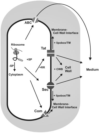
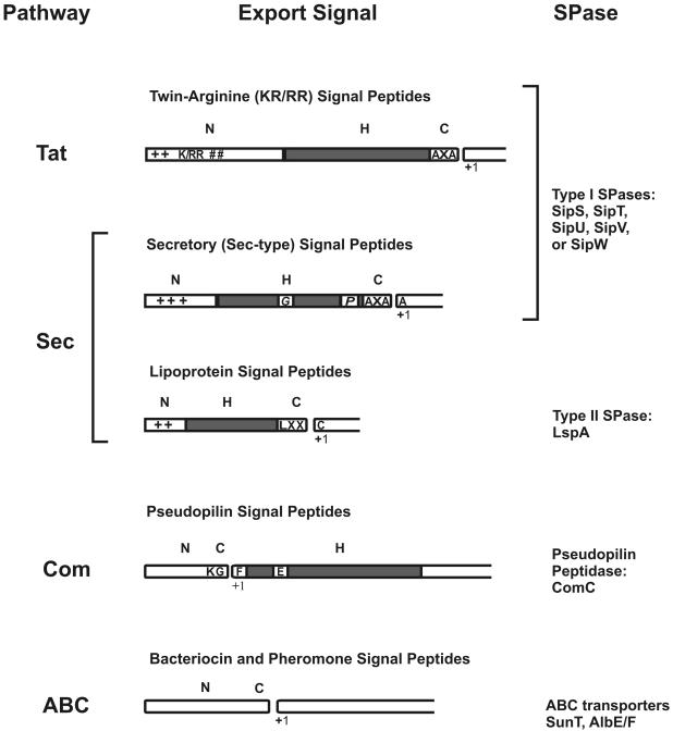
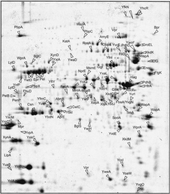
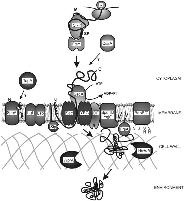
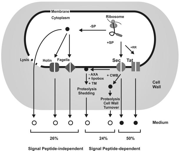
Secretory proteins perform a variety of important "remote-control" functions for bacterial survival in the environment. The availability of complete genome sequences has allowed us to make predictions about the composition of bacterial machinery for protein secretion as well as the extracellular complement of bacterial proteomes. Recently, the power of proteomics was successfully employed to evaluate genome-based models of these so-called secretomes. Progress in this field is well illustrated by the proteomic analysis of protein secretion by the gram-positive bacterium Bacillus subtilis, for which approximately 90 extracellular proteins were identified. Analysis of these proteins disclosed various "secrets of the secretome," such as the residence of cytoplasmic and predicted cell envelope proteins in the extracellular proteome. This showed that genome-based predictions reflect only approximately 50% of the actual composition of the extracellular proteome of B. subtilis. Importantly, proteomics allowed the first verification of the impact of individual secretion machinery components on the total flow of proteins from the cytoplasm to the extracellular environment. In conclusion, proteomics has yielded a variety of novel leads for the analysis of protein traffic in B. subtilis and other gram-positive bacteria. Ultimately, such leads will serve to increase our understanding of virulence factor biogenesis in gram-positive pathogens, which is likely to be of high medical relevance.


Figures

FIG. 1.
FIG. 1.
Protein export pathways in B. subtilis. Ribosomally synthesized proteins can be sorted to various destinations depending on the presence (+SP) or absence (−SP) of an N-terminal signal peptide and specific retention signals. Proteins devoid of a signal peptide remain in the cytoplasm. Proteins that have to be retained at the extracytoplasmic side of the membrane can contain either a transmembrane segment (TM) or a lipid modification (+lipobox). They are exported via the Sec or Tat pathway. Pseudopilins are exported by the Com system. Proteins that need to be retained in the cell wall can be exported via either the Sec or Tat pathway. To be retained in the cell wall, the mature parts of these proteins contain cell wall-binding repeats (+CWB). Proteins can be secreted into the medium via the Sec or Tat pathway or by ABC transporters.
FIG. 2.
FIG. 2.
Classification of cleavable N-terminal signal peptides. On the basis of SPase cleavage sites and the export pathways via which the preproteins are exported, predicted signal peptides (129) were divided into five distinct classes: twin-arginine (RR/KR) signal peptides, secretory (Sec-type) signal peptides, lipoprotein signal peptides, pseudopilin-like signal peptides, and bacteriocin and pheromone signal peptides. The export pathways via which the preproteins are exported and the SPases responsible for their cleavage are indicated. Most signal peptides have a tripartite structure: a positively charged N-domain (N), containing lysine and/or arginine residues (indicated by +), a hydrophobic H-domain (H, indicated by a gray box), and a C-domain (C) that specifies the cleavage site for their specific SPase. The length of the signal peptides and their subdomains is drawn to the same scale. Furthermore, helix-breaking residues, mostly glycine or proline (G/P), in the H-domain of Sec-type signal peptides are indicated. These residues are, respectively, thought to facilitate loopwise membrane insertion and cleavage by SPase I (129). Finally, where appropriate, the most frequently occurring first amino acid of the mature protein (+1) is indicated.
FIG. 3.
FIG. 3.
Master gel for the extracellular proteome of B. subtilis 168. Cells of B. subtilis 168 were grown in LB broth, and extracellular proteins were harvested 1 h after entry into the stationary growth phase. After precipitation with trichloroacetic acid, the extracellular proteins were separated by 2D PAGE and stained with Sypro Ruby as described by Jongbloed et al. (51). The proteins identified by mass spectrometry and/or N-terminal amino acid sequencing are indicated on the gel and listed in Table 1. The extracellular proteins found specifically during growth in minimal media (i.e., YdhT, YflE, and GapA) (46) and those specifically found during growth in phosphate starvation medium (i.e., GlpQ, PhoA, PhoB, PhoD, PstS, YdhF, YcdH, and YrpE) (5, 6, 51, 52) cannot be seen on this gel.
FIG. 4.
FIG. 4.
Components involved in Sec-dependent protein export in B. subtilis. Secretory proteins are ribosomally synthesized as precursor proteins with an N-terminal signal peptide (SP). Cytoplasmic chaperones, such as SRP/FtsY (47) and CsaA (75, 76), keep the precursors in a translocation-competent state and facilitate their targeting to the translocase in the membrane, consisting of SecA, SecY, SecE, SecG, and SecDF (17, 129). During or shortly after translocation, the preprotein is cleaved by one of the type I signal peptidases (SipS-W) (130) or lipid modified by the diacylglyceryl-transferase (Lgt) (62) and cleaved by the lipoprotein-specific signal peptidase (Lsp) (136). SppA and TepA may be involved in the degradation of cleaved signal peptides (16), whereas the folding of several secreted proteins depends on the activities of PrsA (55), BdbBC (18), and/or SpoIIIJ/YqjG (135). HtrA, HtrB (85), and WprA (68, 124) are involved in the quality control of secretory proteins. It should be noted that for reasons of simplicity, HtrAB are depicted in the cell wall, although HtrA is detected in both the membrane and the medium (5). On passage through the cell wall, the mature protein is released into the environment.
FIG. 5.
FIG. 5.
Mechanisms of extracellular accumulation of B. subtilis proteins. Ribosomally synthesized proteins can be sorted to various destinations, depending on the presence (+SP) or absence (−SP) of an N-terminal signal peptide and specific retention signals. Based on the results obtained by proteomic studies, about 50% of the extracellular proteome is directly secreted into the medium via the Sec and Tat pathways. Notably, only one protein, PhoD, is (so far) known to be secreted via the Tat pathway. Proteins which have to be retained at the extracytoplasmatic side of the membrane can either lack an SPase cleavage site (−AXA), be lipid-modified (+lipobox), contain transmembrane (+TM) domains, or contain cell wall-binding repeats (+CWB). Such retained proteins are exported from the cytoplasm via the Sec or Tat pathways. About 24% of the proteins found on the extracellular proteome are predicted to have retention signals. These proteins are released into the medium by proteolysis, shedding, or cell wall turnover. Finally, about 26% of the extracellular proteome lacks typical signal peptides and can escape from the cytoplasm by cell lysis or via the flagellar export machinery, the holin systems, or other unidentified export systems. Expected locations of identified proteins, as based on previous genome-based predictions (129), are indicated by solid circles, whereas unexpected locations of identified proteins are indicated by open circles.
FIG. 6.
FIG. 6.
Average features of 56 identified Sec-type signal peptides. Average signal peptide length and the length and hydrophobicity of the N- and H domains were determined on the basis of the Sec-type signal peptides from 52 identified extracellular proteins (Table 1) (5, 6, 129) and 4 cell wall-located proteins (Table 3); (6) with (putative) SPase I cleavage sites. The YfnI and YflE proteins are excluded from the analysis because they contain N-terminal transmembrane domains rather than typical signal peptides. We have included all cleavable signal peptides with RR/KR motifs, exept that of PhoD, since the export of the corresponding proteins was shown to be Tat independent (51, 52). aa, amino acids.
FIG. 7.
FIG. 7.
Relative contributions of different mechanisms for extracellular protein accumulation in gram-positive bacteria. The relative contribution of different mechanisms for the extracellular accumulation of proteins from gram-positive bacteria was deduced from extracellular proteome studies with B. subtilis, B. cereus, C. difficile, S. aureus, GAS, and M. tuberculosis (see the text for details). For this overview, the number of proteins released by a certain export mechanism was devided by the total number of identified extracellular proteins. Sec, signal peptide- and Sec-dependent protein secretion; Lys, typical cytoplasmic proteins released by lysis or an unidentified signal peptide-independent mechanism; Hol, release of proteins by specific holin systems, Fla, release of flagellum-related proteins. It should be noted that the Sec portion of the extracellular proteomes includes all proteins with Sec-type signals, lipoprotein signals, and transmembrane domains that have the potential to direct transport across the membrane. Because only one protein of B. subtilis (PhoD) is known to be secreted in a Tat-dependent manner, this group of proteins was not included in this comparison.

References

    1. Akita, M., S. Sasaki, S. Matsuyama, and S. Mizushima. 1990. SecA interacts with secretory proteins by recognizing the positive charge at the amino terminus of the signal peptide in Escherichia coli. J. Biol. Chem. 265:8162-8169. - PubMed
    1. Alami, M., I. Lüke, S. Deitermann, J. Brunner, and M. Müller. 2003. Differential interactions between a twin-arginine signal peptide and its translocase in Escherichia coli. Mol. Cell 12:937-946. - PubMed
    1. Antelmann, H., C. Scharf, and M. Hecker. 2000. Phosphate starvation-inducible proteins of Bacillus subtilis: proteomics and transcriptional analysis. J. Bacteriol. 182:4478-4490. - PMC - PubMed
    1. Antelmann, H., E. Darmon, D. Noone, J. W. Veening, H. Westers, S. Bron, O. P. Kuipers, K. M. Devine, M. Hecker, and J. M. van Dijl. 2003. The extracellular proteome of Bacillus subtilis under secretion stress conditions. Mol. Microbiol. 49:143-156. - PubMed
    1. Antelmann, H., H. Tjalsma, B. Voigt, S. Ohlmeier, S. Bron, J. M. van Dijl, and M. Hecker. 2001. A proteomic view on genome-based signal peptide predictions. Genome Res. 11:14984-14502. - PubMed

Publication types

MeSH terms

LinkOut - more resources