Review
. 2004 Jun;68(2):234-62.
doi: 10.1128/MMBR.68.2.234-262.2004.

Compartmentalization of gene expression during Bacillus subtilis spore formation

Affiliations
Review

Compartmentalization of gene expression during Bacillus subtilis spore formation

David W Hilbert et al. Microbiol Mol Biol Rev. 2004 Jun.

Abstract

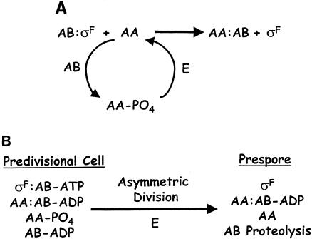
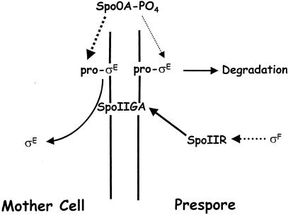
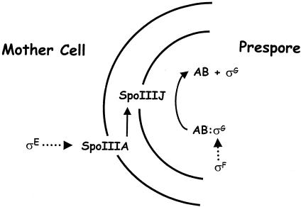
Gene expression in members of the family Bacillaceae becomes compartmentalized after the distinctive, asymmetrically located sporulation division. It involves complete compartmentalization of the activities of sporulation-specific sigma factors, sigma(F) in the prespore and then sigma(E) in the mother cell, and then later, following engulfment, sigma(G) in the prespore and then sigma(K) in the mother cell. The coupling of the activation of sigma(F) to septation and sigma(G) to engulfment is clear; the mechanisms are not. The sigma factors provide the bare framework of compartment-specific gene expression. Within each sigma regulon are several temporal classes of genes, and for key regulators, timing is critical. There are also complex intercompartmental regulatory signals. The determinants for sigma(F) regulation are assembled before septation, but activation follows septation. Reversal of the anti-sigma(F) activity of SpoIIAB is critical. Only the origin-proximal 30% of a chromosome is present in the prespore when first formed; it takes approximately 15 min for the rest to be transferred. This transient genetic asymmetry is important for prespore-specific sigma(F) activation. Activation of sigma(E) requires sigma(F) activity and occurs by cleavage of a prosequence. It must occur rapidly to prevent the formation of a second septum. sigma(G) is formed only in the prespore. SpoIIAB can block sigma(G) activity, but SpoIIAB control does not explain why sigma(G) is activated only after engulfment. There is mother cell-specific excision of an insertion element in sigK and sigma(E)-directed transcription of sigK, which encodes pro-sigma(K). Activation requires removal of the prosequence following a sigma(G)-directed signal from the prespore.


Figures

FIG. 1.
FIG. 1.
Schematic representation of the stages of spore formation. A vegetatively growing cell is defined as stage 0. It is shown as having completed DNA replication and containing two complete chromosomes (represented as disordered lines within the cells), although replication is not completed at the start of spore formation. Formation of an axial filament of chromatin, where both chromosomes (or a partially replicated chromosome) form a continuous structure that stretches across the long axis of the cell, is defined as stage I. Asymmetric division occurs at stage II, dividing the cell into the larger mother cell and smaller prespore; for clarity, the septum is indicated as a single line. At the time of division, only approximately 30% of a chromosome is trapped in the prespore, but the DNA translocase SpoIIIE will rapidly pump in the remaining 70%. Stage III is defined as completion of engulfment, and the prespore now exists as a free-floating protoplast within the mother cell enveloped by two membranes, represented by a single ellipse. Synthesis of the primordial germ cell wall and cortex, a distinctive form of peptidoglycan, between the membranes surrounding the prespore is defined as stage IV and is represented as thickening and graying of the ellipse. Deposition of the spore coat, protective layers of proteins around the prespore, is defined as stage V. The coat is represented as the black layer surrounding the engulfed prespore. Coincident with coat and cortex formation, the engulfed prespore is dehydrated, giving it a phase-bright appearance, represented here as a light grey shading. Stage VI is maturation, when the spore acquires its full resistance properties, although no obvious morphological changes occur. Stage VII represents lysis of the mother cell, which releases the mature spore into the environment.
FIG. 2.
FIG. 2.
Intercompartmental communication during sporulation. The parallel vertical lines represent the two membranes separating the prespore (right) from the mother cell (left). Diagonal red lines represent pathways of intercompartmental posttranslational activation, and vertical black arrows represent intracompartmental transcriptional activation. Fluorescent micrographs represent cells stained with FM4-64 (red) to visualize the cell membranes and expressing compartment-specific gfp fusions to spoIIQ, spoIID, sspA, and gerE for σF, σE, σG, and σK, respectively. The prespore membranes are not stained in the σG and σK images because engulfment is complete and the prespore membranes are now inaccessible to the lipophilic FM4-64 stain. σF, active in the prespore, is the first compartmentalized σ factor during sporulation. It triggers expression of SpoIIR, which activates the inferred receptor protease SpoIIGA, located in the asymmetric septum. Upon receipt of the signal, SpoIIGA processes the inactive precursor pro-σE into active σE in the mother cell. RNA polymerase with σE transcribes the spoIIIA operon, whose products then signal across the prespore membrane to activate σG, expressed in the prespore under the control of σF but held inactive by SpoIIAB (and probably other factors) until this signal is received. SpoIIIJ is also required for this signaling; although only required (and therefore only represented) in the prespore, it is expressed vegetatively and is presumably present in both compartments. Although not represented here, transcription of spoIIIG (encoding σG) requires an unknown signal from the mother cell as well as the SpoIIQ protein, expressed in the prespore under the control of σF. Once σG becomes active, it causes expression of SpoIVB, which is inserted into the inner prespore membrane. SpoIVB triggers processing of pro-σK, which is synthesized in the mother cell from the σE-directed sigK gene. The processing enzyme is thought to be SpoIVFB, which also expressed in the mother cell under the control of σE but does not act upon pro-σK until it receives the SpoIVB signal from the prespore.
FIG. 3.
FIG. 3.
Chromosome partitioning and genetic asymmetry. A single cell progressing through sporulation is represented on the right. Disordered internal lines represent the nucleoids. The earliest (topmost) cell is drawn as having two complete chromosomes, although it may contain one partially replicated chromosome. Through the action of DivIVA, RacA, and Soj, the two complete chromosomes (or the partially replicated chromosome) are remodeled into an axial filament that extends across the long axis of the cell, represented in the second cell. After asymmetric division occurs, the prespore contains only the origin-proximal one-third of a chromosome, whereas the mother cell contains one complete chromosome and two-thirds of another; this partitioning results in transient genetic asymmetry between the mother cell and the prespore. For simplicity, the septum is represented as a single line. A portion of the third cell has been expanded in order to represent the asymmetry more clearly; the hatched ovals represent the DNA translocase SpoIIIE; the locations of several genetic loci are noted; and σF is depicted as being active in the prespore and σE is depicted as being active in the mother cell. Within about 15 min of the asymmetric division, SpoIIIE pumps the remaining two-thirds of the prespore chromosome into this compartment, restoring genetic symmetry.
FIG. 4.
FIG. 4.
Models of σF regulation. AA, AB, and E refer to SpoIIAA, SpoIIAB, and SpoIIE, respectively. The anti-σ factor SpoIIAB binds σF as a dimer but is represented here as a monomer for simplicity. (A)Basic model of σF regulation. The anti-σ factor SpoIIAB binds σF and holds it inactive. This inhibition can be reversed by the anti-anti-σ factor SpoIIAA. SpoIIAA is subject to regulation by its phosphorylation state; it is inactive when phosphorylated by SpoIIAB (a serine kinase as well as an anti-σ factor) and active when dephosphorylated by SpoIIE. Once dephosphorylated, SpoIIAA can bind SpoIIAB and liberate σF, activating prespore-specific transcription. In this model, the phosphorylation state of SpoIIAA is directly correlated with σF activity, and the fate of SpoIIAB after σF liberation and the nucleotide binding status of SpoIIAB are not considered. (B) Integrated model of σF regulation. In the predivisional cell, SpoIIAA and SpoIIAB are present in two forms: phosphorylation of SpoIIAA by SpoIIAB results in free phosphorylated (inactive) SpoIIAA and a SpoIIAA-SpoIIAB-ADP complex, while unreacted SpoIIAB-ATP forms an inhibitory complex with σF. As long as the level of dephosphorylated SpoIIAA remains below a certain threshold, it will be absorbed by the SpoIIAB-ADP sink. Asymmetric division triggers activation of σF in the prespore through three possible mechanisms: generation of excess dephosphorylated SpoIIAA so that the sink can no longer absorb all of it, sequestration of SpoIIAB in a long-lived complex with SpoIIAA, and proteolysis of SpoIIAB. Asymmetric division is thought to increase the level of dephosphorylated SpoIIAA either by activation of the phosphatase activity of SpoIIE or equivalent distribution of SpoIIE into both compartments, resulting in a much higher SpoIIE/SpoIIAA-PO4 ratio in the prespore. The complexes listed are not intended to reflect a stoichiometric biochemical reaction; rather, they reflect the different combinations thought to be formed by these factors and how they correlate with asymmetric division and activation of σF. The mother cell (not shown) is presumed to resemble the predivisional cell.
FIG. 5.
FIG. 5.
Regulation of σE activation. Parallel vertical lines separating the prespore (right) from the mother cell (left) represent the asymmetric septum. Broken arrows represent transcriptional activation, and solid arrows represent posttranslational regulation. Spo0A-PO4 is present in the predivisional cell as well as both compartments and therefore is represented above the sporulation septum. Pro-σE is synthesized in a Spo0A-PO4-dependent manner and therefore is present in both compartments; however, recent study has indicated that Spo0A-PO4-dependent transcription is largely confined to the mother cell after asymmetric division. This distinction is represented here by a thick line, indicating a high level of expression, in the mother cell and a thin line, indicating a low level of expression, in the prespore. Pro-σE, which is membrane bound, is processed into the active form, σE, by the inferred membrane-bound protease SpoIIGA. SpoIIGA becomes active in response to SpoIIR, whose expression is activated by σF. SpoIIGA is presumably present in both compartments, but σE becomes active only in the mother cell, at least in part because of the higher concentration of pro-σE in this compartment, as well as because of degradation of pro-σE in the prespore. The prespore specificity of SpoIIR expression may contribute to but is not critical for mother cell-specific activation of σE.
FIG. 6.
FIG. 6.
Regulation of σG activation. Two concentric semicircles represent the inner and outer membranes of the engulfed prespore. Broken arrows represent transcriptional activation, and solid arrows represent posttranslational regulation. The structural gene for σG, spoIIIG, is transcribed exclusively in the prespore under the control of σF (SpoIIAB [AB] is presumably inherited from the predivisional cell). Although not represented here, transcription also requires expression of SpoIIQ in the prespore and an unknown gene in the mother cell. Activation of σG does not occur until engulfment is complete. Activation requires release of inhibition by SpoIIAB. This release depends on the σE-directed expression of the spoIIIA operon in the mother cell. Activation of σG also requires SpoIIIJ, which is expressed vegetatively and localizes to the prespore membrane but need only be expressed in the prespore. It seems likely that some mechanism distinct from SpoIIAB inhibition keeps σG inactive prior to engulfment. The mechanism responsible for coupling activation to completion of engulfment remains unclear.
FIG. 7.
FIG. 7.
Regulation of σK activation. Two concentric semicircles indicate the inner and outer prespore membranes surrounding the engulfed prespore. Broken arrows represent transcriptional activation, and solid arrows represent posttranslational regulation. SpoIVB is expressed in the prespore under the control of σG and is thought to be inserted into the inner prespore membrane, where it undergoes autoproteolysis. σG also directs expression of BofC, which is an inhibitor of SpoIVB. In the mother cell, BofA, SpoIVFA, SpoIVFB, and pro-σK are all produced under the control of σE. SpoIVFB is thought to be the processing enzyme that acts upon pro-σK to generate active σK. BofA inhibits SpoIVFB. This inhibition is mediated by SpoIVFA, which acts to bring these proteins in contact with one another. Signaling by SpoIVB relieves inhibition of SpoIVFB, possibly by proteolysis, and so triggers pro-σK processing. Pro-σK is tethered to the outer prespore membrane by its N terminus, and it is thought that the processing reaction with SpoIVFB occurs within the membrane.

Similar articles

Cited by

References

    1. Abanes-De Mello, A., Y. L. Sun, S. Aung, and K. Pogliano. 2002. A cytoskeleton-like role for the bacterial cell wall during engulfment of the Bacillus subtilis forespore. Genes Dev. 16:3253-3264. - PMC - PubMed
    1. Adams, L. F., K. L. Brown, and H. R. Whiteley. 1991. Molecular cloning and characterization of two genes encoding σ factors that direct transcription from a Bacillus thuringiensis crystal protein gene promoter. J. Bacteriol. 173:3846-3854. - PMC - PubMed
    1. Alper, S., L. Duncan, and R. Losick. 1994. An adenosine nucleotide switch controlling the activity of a cell type-specific transcription factor in B. subtilis. Cell 77:195-205. - PubMed
    1. Amaya, E., A. Khvorova, and P. J. Piggot. 2001. Analysis of promoter recognition in vivo directed by σF of Bacillus subtilis by using random-sequence oligonucleotides. J. Bacteriol. 183:3623-3630. - PMC - PubMed
    1. Andreoli, A. J., S. Suehiro, D. Sakiyama, J. Takemoto, E. Vivanco, J. C. Lara, and M. C. Klute. 1973. Release and recovery of forespores from Bacillus cereus. J. Bacteriol. 115:1159-1166. - PMC - PubMed

Publication types

LinkOut - more resources