Review
. 2004 Jun;68(2):301-19.
doi: 10.1128/MMBR.68.2.301-319.2004.

Diversity in chemotaxis mechanisms among the bacteria and archaea

Affiliations
Review

Diversity in chemotaxis mechanisms among the bacteria and archaea

Hendrik Szurmant et al. Microbiol Mol Biol Rev. 2004 Jun.

Abstract

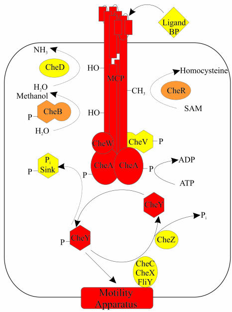
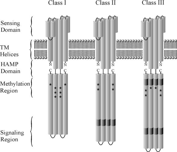
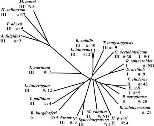
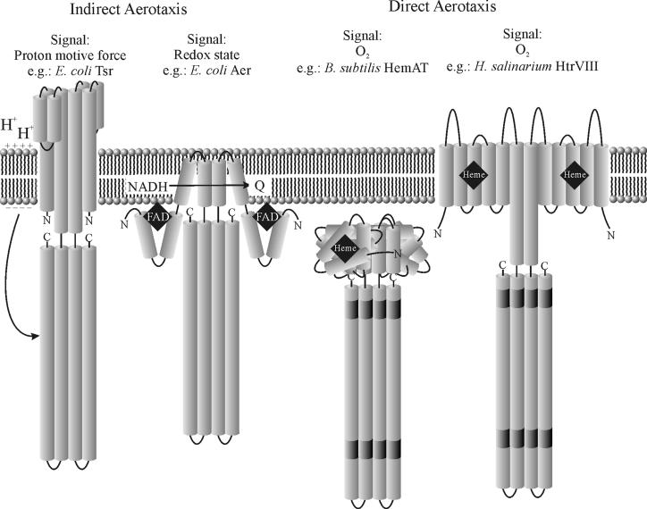
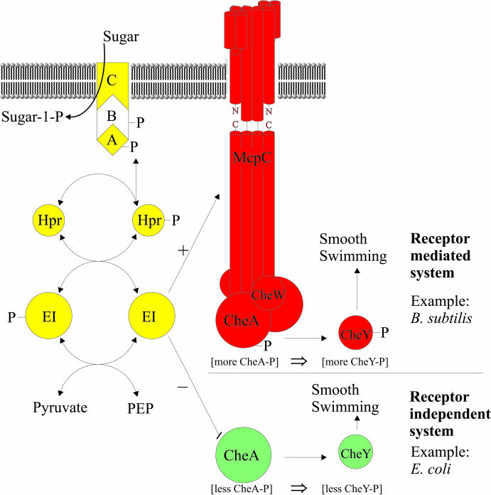
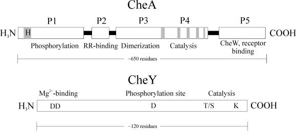
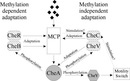
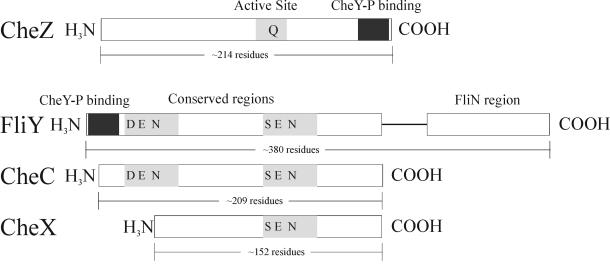
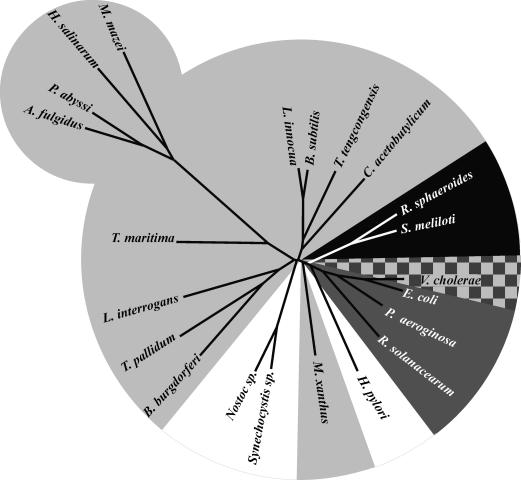
The study of chemotaxis describes the cellular processes that control the movement of organisms toward favorable environments. In bacteria and archaea, motility is controlled by a two-component system involving a histidine kinase that senses the environment and a response regulator, a very common type of signal transduction in prokaryotes. Most insights into the processes involved have come from studies of Escherichia coli over the last three decades. However, in the last 10 years, with the sequencing of many prokaryotic genomes, it has become clear that E. coli represents a streamlined example of bacterial chemotaxis. While general features of excitation remain conserved among bacteria and archaea, specific features, such as adaptational processes and hydrolysis of the intracellular signal CheY-P, are quite diverse. The Bacillus subtilis chemotaxis system is considerably more complex and appears to be similar to the one that existed when the bacteria and archaea separated during evolution, so that understanding this mechanism should provide insight into the variety of mechanisms used today by the broad sweep of chemotactic bacteria and archaea. However, processes even beyond those used in E. coli and B. subtilis have been discovered in other organisms. This review emphasizes those used by B. subtilis and these other organisms but also gives an account of the mechanism in E. coli.


Figures

FIG. 1.
FIG. 1.
General chemotaxis model. A schematic of the biochemical processes in the two-component chemotaxis pathway is shown. Hexagons represent response regulator domains. The universal components are in red; almost universal components are in orange; optional components are in yellow.
FIG. 2.
FIG. 2.
Schematic of the three classes of chemotaxis receptors. Shown is a representative dimer for each class of chemotaxis receptors. Insertion/deletion regions (indels) are shaded in dark gray. Stars indicate sites of methylation for (from left to right) E. coli Tar, M. xanthus FrzCD, and B. subtilis McpB.
FIG. 3.
FIG. 3.
Phylogenetic tree of chemotactic bacteria and archaea. The phylogenetic tree was generated from the 16S rRNA sequences by using the programs CLUSTALW and DRAWTREE. Included is information regarding the number and class of chemoreceptors for each respective organism. ND, not determined. Organisms, by phylum, are as follows: Archaea: Archaeoglobus fulgidus, Halobacterium salinarum, Methanosarcina mazei, and Pyrococcus abyssi; Thermotogales: Thermotoga maritima; spirochetes: Borrelia burgdorferi, Leptospira interrogans, and Treponema pallidum; cyanobacteria: Nostoc and Synechocystis spp.; gram-positive bacteria: Bacillus subtilis, Clostridium acetobutylicum, Listeria innocua, and Thermoanaerobacter tengcongensis; proteobacteria (α-subgroup): Rhodobacter sphaeroides and Sinorizhobium meliloti; proteobacteria (β-subgroup): Ralstonia solanacearum; proteobacteria (δ-subgroup): Myxococcus xanthus; proteobacteria (ɛ-subgroup): Helicobacter pylori; proteobacteria (γ-subgroup): Escherichia coli, Pseudomonas aeroginosa, and Vibrio cholerae.
FIG. 4.
FIG. 4.
Aerotaxis receptors. Shown is a schematic of the four known types of aerotaxis receptors. Indirect aerotaxis defines receptors that detect oxygen levels by the proton motive force or redox state of the cell. Direct aerotaxis defines receptors that detect levels directly by interacting with oxygen. Black diamonds represent the indicated receptor cofactors.
FIG. 5.
FIG. 5.
PTS in chemotaxis. The two known chemotaxis pathways for PTS sugars are shown. Transport of PTS sugars increases the concentration of unphosphorylated enzyme I (EI) that can either directly interact and inhibit CheA (receptor-independent system), as is the case in E. coli, or indirectly stimulate CheA through the receptors (receptor-dependent system), as thought to be the case in B. subtilis. The letters A, B, and C represent components of an ABC transporter. For better understanding, PTS proteins are in yellow, B. subtilis chemotaxis proteins are in red, and E. coli chemotaxis proteins are in green.
FIG. 6.
FIG. 6.
Schematic of CheA and CheY. Shown are the histidine kinase CheA and the general response regulator CheY. For CheA, the five domains are labeled P1 through P5. The phosphoreceiving histidine in P1 is highlighted in gray, and conserved regions within domain P4 that are thought to play an active role in catalysis are also highlighted in gray. For CheY, conserved residues that participate in catalysis are positioned as indicated.
FIG. 7.
FIG. 7.
Adaptation systems. A flowchart of the possible means of adaptation is shown. The almost universal methylation-dependent adaptation system is shown on the left. The less highly conserved methylation-independent pathways are on the right. We speculate here that CheV might directly influence CheA activity following phosphorylation, as shown, while the means of CheC activation and adaptational action are not yet understood. Black diamonds represent a chemoeffector. Adaptational proteins are hatched; excitatory proteins are shaded.
FIG. 8.
FIG. 8.
Schematic of CheY-P-hydrolyzing proteins. For CheZ, the C-terminal CheY-P binding region is shown in black and the area including what is thought to be the active site is shaded in gray. For FliY, the CheY-P binding site is shown in black. For FliY, CheC, and CheX, conserved regions are in gray, with highly conserved residues positioned as indicated.
FIG. 9.
FIG. 9.
Means of CheY-P hydrolysis in chemotactic organisms. The tree was generated as described for Fig. 3. Chemotactic organisms that encode a CheC homolog are highlighted in light gray; dark gray represents organisms that encode a CheZ; black represents organisms that encode an alternative CheY that acts as a phosphate sink; white represents organisms with no known mechanism of CheY-P hydrolysis. V. cholerae encodes both CheZ and CheC homologs and is therefore indicated by dark and light gray squares.

References

    1. Adler, J., G. L. Hazelbauer, and M. M. Dahl. 1973. Chemotaxis toward sugars in Escherichia coli. J. Bacteriol. 115:824-847. - PMC - PubMed
    1. Aizawa, S. I., Zhulin, I. B., L. Marquez-Magana, and G. W. Ordal. 2002. Chemotaxis and motility, p. 437-452. In A. L. Sonenshein, R. Losick, and J. A. Hoch (ed.), Bacillus subtilis and its closest relatives: from genes to cells. ASM Press, Washington, D.C.
    1. Aksamit, R., and D. E. Koshland, Jr. 1972. A ribose binding protein of Salmonella typhimurium. Biochem. Biophys. Res. Commun. 48:1348-1353. - PubMed
    1. Aksamit, R. R., and D. E. Koshland, Jr. 1974. Identification of the ribose binding protein as the receptor for ribose chemotaxis in Salmonella typhimurium. Biochemistry 13:4473-4478. - PubMed
    1. Alam, M., M. Claviez, D. Oesterhelt, and M. Kessel. 1984. Flagella and motility behaviour of square bacteria. EMBO J. 3:2899-2903. - PMC - PubMed

Publication types

LinkOut - more resources