Review
. 2004 Jun;68(2):345-61.
doi: 10.1128/MMBR.68.2.345-361.2004.

Microbial responses to microgravity and other low-shear environments

Collaborators, Affiliations
Review

Microbial responses to microgravity and other low-shear environments

Cheryl A Nickerson et al. Microbiol Mol Biol Rev. 2004 Jun.

Abstract

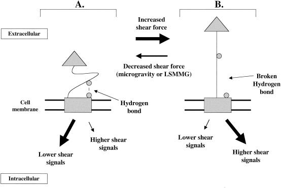
Microbial adaptation to environmental stimuli is essential for survival. While several of these stimuli have been studied in detail, recent studies have demonstrated an important role for a novel environmental parameter in which microgravity and the low fluid shear dynamics associated with microgravity globally regulate microbial gene expression, physiology, and pathogenesis. In addition to analyzing fundamental questions about microbial responses to spaceflight, these studies have demonstrated important applications for microbial responses to a ground-based, low-shear stress environment similar to that encountered during spaceflight. Moreover, the low-shear growth environment sensed by microbes during microgravity of spaceflight and during ground-based microgravity analogue culture is relevant to those encountered during their natural life cycles on Earth. While no mechanism has been clearly defined to explain how the mechanical force of fluid shear transmits intracellular signals to microbial cells at the molecular level, the fact that cross talk exists between microbial signal transduction systems holds intriguing possibilities that future studies might reveal common mechanotransduction themes between these systems and those used to sense and respond to low-shear stress and changes in gravitation forces. The study of microbial mechanotransduction may identify common conserved mechanisms used by cells to perceive changes in mechanical and/or physical forces, and it has the potential to provide valuable insight for understanding mechanosensing mechanisms in higher organisms. This review summarizes recent and future research trends aimed at understanding the dynamic effects of changes in the mechanical forces that occur in microgravity and other low-shear environments on a wide variety of important microbial parameters.


Figures

FIG. 1.
FIG. 1.
Operating orientations of the RWV and the effect of RWV rotation on particle (microbe) suspension. (A) The two operating orientations of the RWV are depicted. In the LSMMG orientation (panel i), the axis of rotation of the RWV is perpendicular to the direction of the gravity force vector. In the normal-gravity (or 1 × g) orientation (panel ii), the axis of rotation is parallel to the gravity vector. (B) Effect of RWV rotation on particle suspension. When the RWV is not rotating or is rotating in the 1 × g orientation (panel i), the force of gravity will cause particles in the apparatus to sediment and eventually settle on the bottom of the RWV. When the RWV is rotating in the LSMMG position (panel ii), particles are continually suspended in the medium. The medium within the RWV rotates as a single body, and the sedimentation of the particle due to gravity is offset by the upward forces of rotation. The result is a low-shear aqueous suspension that is strikingly similar to what would occur in true microgravity. Panel B is not drawn to scale.
FIG. 2.
FIG. 2.
LSMMG-enhanced virulence of S. enterica serovar Typhimurium in the murine model of infection. (A) Shortened time to death of mice infected with LSMMG-grown S. enterica serovar Typhimurium cells compared to mice infected with 1 × g-grown cells. BALB/c mice (8 weeks old) were inoculated perorally with 2 × 10⁶ CFU of LSMMG- or 1 × g-grown S. enterica serovar Typhimurium, and the survival of the animals was monitored for 20 days postinfection. The percent survival of the infected animals over this period is plotted, and the curves for LSMMG- and 1 × g-infected mice are indicated. (B) Enhanced ability of LSMMG-grown S. enterica serovar Typhimurium cells to colonize the murine spleen and liver compared to that of 1 × g-grown cells. LSMMG- and 1 × g-grown S. enterica serovar Typhimurium cells (2 × 10⁶) were administered perorally as individual infections to 8-week-old BALB/c mice. The spleen and liver were excised 6 days after infection, and the recovered bacteria were quantitated. The standard deviation represents the statistical difference between five mice for each infection group. (Reprinted from reference .)
FIG. 3.
FIG. 3.
Chromosomal organization of the S. enterica serovar Typhimurium LSMMG regulon. The circular chromosome is schematically depicted, with kilobase coordinates noted and labeled. LSMMG-regulated genes as identified by microarray analysis are noted as unlabeled lines extending from the chromosome. The genes belong to diverse functional groups including transcriptional regulators, virulence factors, LPS biosynthetic enzymes, ribosomal proteins, iron utilization functions, and proteins of unknown function. The numbered brackets indicate clusters of LSMMG-regulated genes that are physically linked (within 50 kb) or part of the same operon. Identification of such physically linked gene clusters is important because they may represent genes that are part of the same “island,” which may contain operons that are coregulated by the same transcriptional regulator. This could give clues to the identity of potential LSMMG regulators. In fact, clusters 7 and 4 contain several LSMMG-responsive genes that belong to the Salmonella pathogenicity islands SPI-1 and SPI-2, respectively. (Reprinted from reference with permission.)
FIG. 4.
FIG. 4.
Diagram summarizing how the LSMMG signal could be transmitted in S. enterica serovar Typhimurium. Based on current data, this diagram depicts a potential picture of LSMMG signal transmission in S. enterica serovar Typhimurium. The mechanical, physical, and/or chemical changes associated with LSMMG culture in the RWV are sensed by the bacterial cell using a hypothetical sensor mechanism. The sensor component of this mechanism (depicted as a rectangle) is probably located at the cell envelope (similar to common prokaryotic response regulator systems), but an intracellular location is possible. Suggested candidates and mechanistic models for this sensor are discussed in the text. This signal is probably transduced to intracellular regulators that regulate the expression of LSMMG-responsive genes. The potential regulators (depicted as cylinders) of the LSMMG response are labeled. Based on experimental data, it appears that the RpoS sigma factor, a logical candidate regulator, is not required for the LSMMG response in S. enterica serovar Typhimurium. Data obtained from microarray-related experiments suggest that the Fur transcriptional regulator is involved in S. enterica serovar Typhimurium LSMMG signal transmission. Given the large number of functional groups of genes regulated by LSMMG, it is likely that other regulators, indicated by the question mark, are also involved. In addition, there may be overlap in the regulation of Fur-regulated LSMMG genes and other such genes controlled by the unknown regulator(s). This potential cross talk is indicated by a double-headed arrow.
FIG. 5.
FIG. 5.
Model for the way in which microbes may sense changes in aqueous shear force and transmit this signal at the molecular level. Since aqueous environments in microgravity and during LSMMG culture in the RWV are associated with lowered shear forces, this model may explain how microbes “sense” these environments. The functioning of the sensor molecule in this model is based on the data for the shear-responsive E. coli FimH adhesin. A membrane-bound protein with two domains connected by a flexible linker is depicted. The rectangular domain is embedded in the membrane and can also serve to initiate signaling inside the cell in response to changes in the linker domain conformation. The triangular domain is extracellular and can initiate changes in linker domain conformation in response to changes in shear stress. (A), Lowered shear. A hydrogen bond (or another type of noncovalent bond) is formed between a residue on the linker domain and a residue on the membrane domain (both depicted as circles) and keeps the linker in a compact conformation. One hydrogen bond is depicted, but several such bonds could exist, involving multiple residues. This compact linker domain conformation causes the membrane domain to respond by activating signaling pathways associated with lower shear and/or deactivating pathways associated with high shear responses. The result would be responses associated with lower shear. (B) Increased shear. The increased shear force sensed by the extracellular domain weakens and breaks the hydrogen bond(s) between the linker domain and the membrane domain. This causes the membrane domain to fire signaling pathways associated with higher shear and decrease signals associated with lowered shear. The result would be responses associated with higher shear. In this model, the microbe would not be responding to changes in gravity force directly but to the lowered aqueous shear force that occurs when a microbe is suspended in an aqueous environment at microgravity or LSMMG.

Similar articles

Cited by

References

    1. Alpatov, A., Y. Il'in, V. Antipov, and M. Tairbekov. 1988. Biological experiments on COSMOS-1887. Kosm. Biol. Aviakosm. Med. 23:26-32. - PubMed
    1. Audia, J. P., C. C. Webb, and J. W. Foster. 2001. Breaking through the acid barrier: an orchestrated response to proton stress by enteric bacteria. Int. J. Med. Microbiol. 291:97-106. - PubMed
    1. Beeson, J. G., S. J. Rogerson, B. M. Cooke, J. C. Reeder, W. Chai, A. M. Lawson, M. E. Molyneux, and G. V. Brown. 2000. Adhesion of Plasmodium falciparum-infected erythrocytes to hyaluronic acid in placental malaria. Nat. Med. 6:86-90. - PMC - PubMed
    1. Berrier, C., M. Besnard, B. Ajouz, A. Coulombe, and A. Ghazi. 1996. Multiple mechanosensitive ion channels from Escherichia coli, activated at different thresholds of applied pressure. J. Membr. Biol. 151:175-187. - PubMed
    1. Berrier, C., A. Coulombe, C. Houssin, and A. Ghazi. 1989. A patch-clamp study of ion channels of inner and outer membranes and of contact zones of E. coli, fused into giant liposomes. Pressure-activated channels are localized in the inner membrane. FEBS Lett. 259:27-32. - PubMed

Publication types

LinkOut - more resources