

Coarctation of the aorta: a secondary cause of hypertension
- PMID: 15187499
- PMCID: PMC8109355
- DOI: 10.1111/j.1524-6175.2004.02868.x
Coarctation of the aorta: a secondary cause of hypertension
Abstract
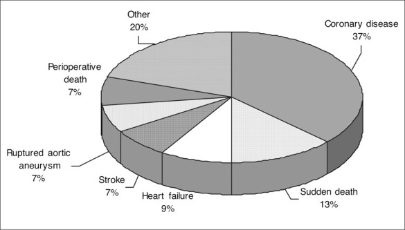
Coarctation of the aorta is a constriction of the aorta located near the ligamentum arteriosum and the origins of the left subclavian artery. This condition may be associated with other congenital disease. The mean age of death for persons with this condition is 34 years if untreated, and is usually due to heart failure, aortic dissection or rupture, endocarditis, endarteritis, cerebral hemorrhage, ischemic heart disease, or concomitant aortic valve disease in uncomplicated cases. Symptoms may not be present in adults. Diminished and delayed pulses in the right femoral artery compared with the right radial or brachial artery are an important clue to the presence of a coarctation of the aorta, as are the presence of a systolic murmur over the anterior chest,bruits over the back, and visible notching of the posterior ribs on a chest x-ray. In many cases a diagnosis can be made with these findings. Two-dimensional echocardiography with Doppler interrogation is used to confirm the diagnosis. Surgical repair and percutaneous intervention are used to repair the coarctation; however, hypertension may not abate. Because late complications including recoarctation, hypertension, aortic aneurysm formation and rupture, sudden death, ischemic heart disease, heart failure, and cerebrovascular accidents may occur, careful follow-up is required.
Figures


Similar articles
-
Discrepancies in aortic growth explain aortic arch gradients during exercise.J Am Coll Cardiol. 1993 Mar 15;21(4):1002-7. doi: 10.1016/0735-1097(93)90360-d. J Am Coll Cardiol. 1993. PMID: 8450148
-
Correction of coarctation of aorta in adult patients--impact of corrective procedure on long-term recoarctation and systolic hypertension.Thorac Cardiovasc Surg. 2008 Mar;56(2):83-6. doi: 10.1055/s-2007-989343. Thorac Cardiovasc Surg. 2008. PMID: 18278682
-
Severe and resistant hypertension in an older woman with claudication.J Am Soc Hypertens. 2017 Aug;11(8):475-479. doi: 10.1016/j.jash.2017.05.007. Epub 2017 Jun 1. J Am Soc Hypertens. 2017. PMID: 28642065 Free PMC article.
-
Coarctation of the aorta in adults: what is the best treatment? Case report and literature review.J Med Life. 2011 May 15;4(2):189-95. Epub 2011 May 25. J Med Life. 2011. PMID: 21776305 Free PMC article. Review.
-
Coarctation of the aorta.Semin Nephrol. 1995 Mar;15(2):87-105. Semin Nephrol. 1995. PMID: 7777727 Review.
Cited by
-
Multi-drug-resistant hypertension caused by severe aortic coarctation presenting in late adulthood.J Clin Hypertens (Greenwich). 2015 Apr;17(4):313-6. doi: 10.1111/jch.12495. Epub 2015 Feb 3. J Clin Hypertens (Greenwich). 2015. PMID: 25644790 Free PMC article.
-
The global effect of aortic coarctation on carotid and renal pulsatile hemodynamics.PLoS One. 2024 Dec 17;19(12):e0310793. doi: 10.1371/journal.pone.0310793. eCollection 2024. PLoS One. 2024. PMID: 39689111 Free PMC article.
-
Novel Approach in Treatment of Coarctation of the Aorta with Bifurcation Stenosis of the Left Subclavian Artery - The Road Less Traveled.Heart Views. 2024 Oct-Dec;25(4):260-263. doi: 10.4103/heartviews.heartviews_64_24. Epub 2025 May 10. Heart Views. 2024. PMID: 40488154 Free PMC article.
-
Hypertensive Heart Disease-The Imaging Perspective.J Clin Med. 2023 Apr 25;12(9):3122. doi: 10.3390/jcm12093122. J Clin Med. 2023. PMID: 37176563 Free PMC article. Review.
-
Emergent Presentation of an Adult with Undiagnosed Coarctation of the Aorta.Case Rep Emerg Med. 2018 Feb 19;2018:5756983. doi: 10.1155/2018/5756983. eCollection 2018. Case Rep Emerg Med. 2018. PMID: 29670774 Free PMC article.
References
-
- Perloff JK. The Clinical Recognition of Congenital Heart Disease. Philadelphia , PA : W.B. Saunders; 2003:113–143.
-
- Ayers CR, Slaughter AR, Smallwood HD, et al. Standards for quality care of hypertensive patients in office and hospital practice. Am J Cardiol. 1973;32:533–545. - PubMed
-
- Rocchini AP. Coarctation of the aorta. In: Izzo JL, Black HR, eds. Hypertension Primer. Dallas , TX : Lippincott Williams & Wilkins; 2003:154–156.
-
- Katira R, Rathore VS, Lip GY, et al. Coarctation of aorta. J Hum Hypertens. 1997;11:537–538. - PubMed
-
- Therrien J, Webb G. Clinical update on adults with congenital heart disease. Lancet. 2003;362:1305–1313. - PubMed
Publication types
MeSH terms
LinkOut - more resources
Full Text Sources
Medical
