. 2004 Jun;3(3):632-45.
doi: 10.1128/EC.3.3.632-645.2004.

The Kip3-like kinesin KipB moves along microtubules and determines spindle position during synchronized mitoses in Aspergillus nidulans hyphae

Affiliations

The Kip3-like kinesin KipB moves along microtubules and determines spindle position during synchronized mitoses in Aspergillus nidulans hyphae

Patricia E Rischitor et al. Eukaryot Cell. 2004 Jun.

Abstract

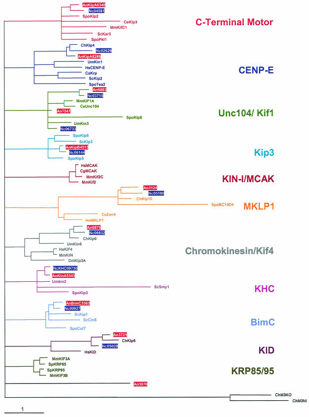
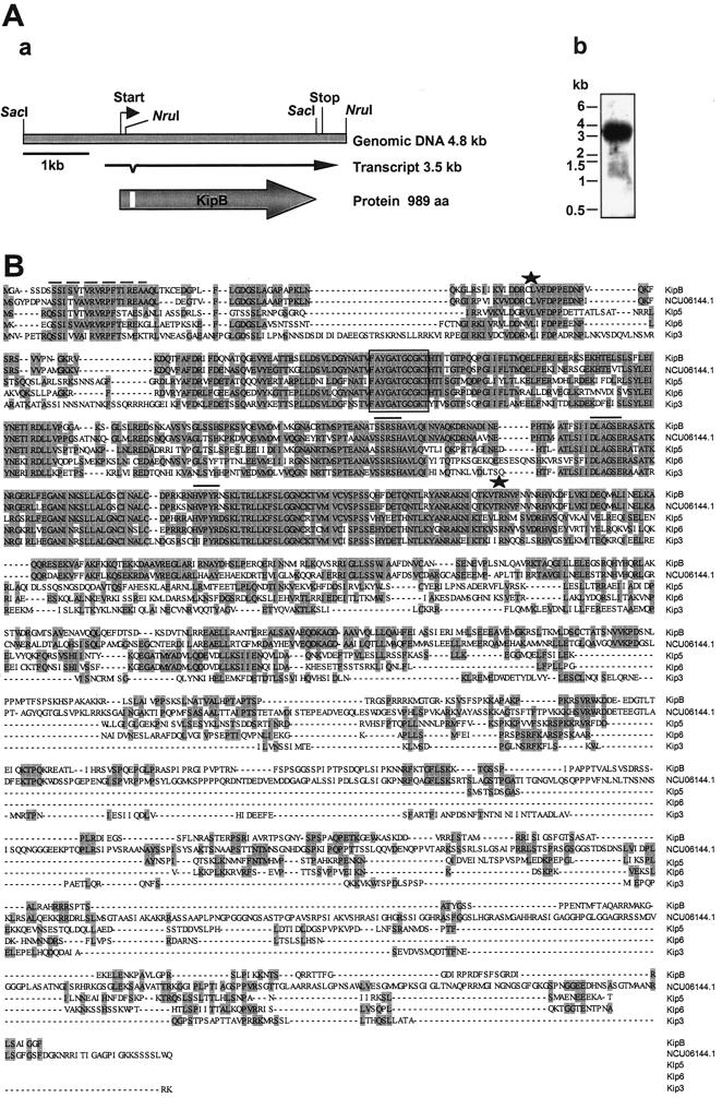
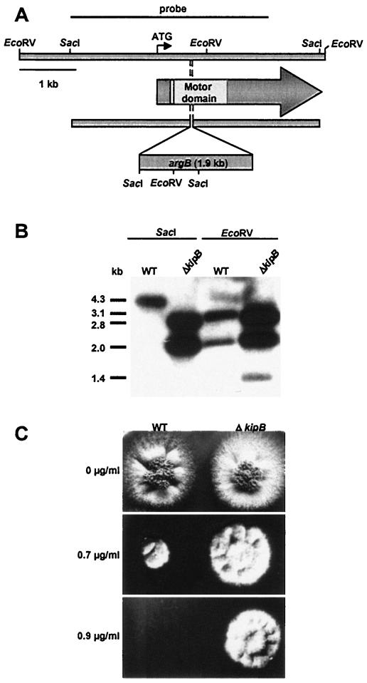
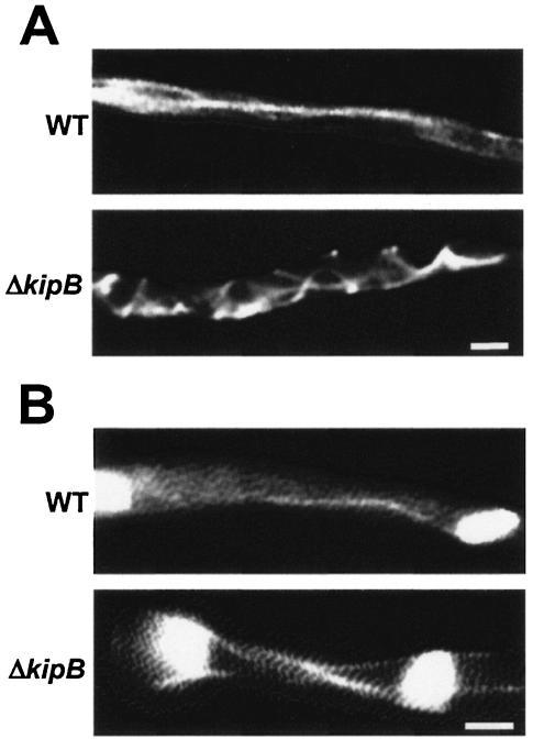
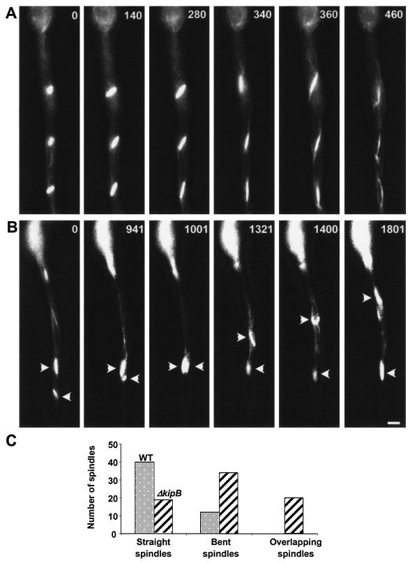
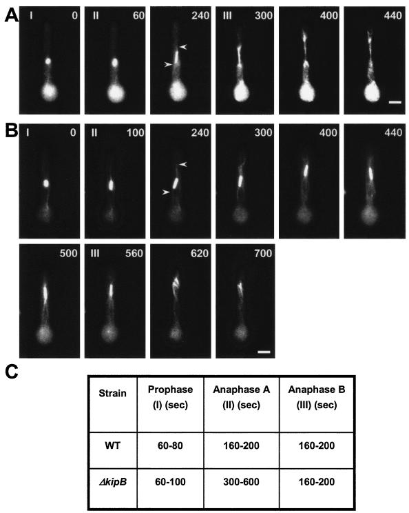
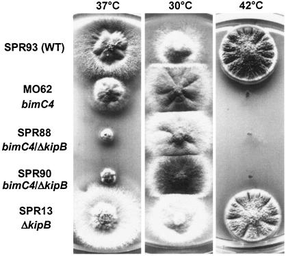
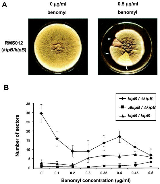
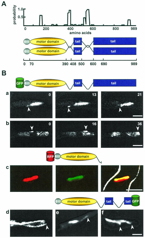
Kinesins are motor proteins which are classified into 11 different families. We identified 11 kinesin-like proteins in the genome of the filamentous fungus Aspergillus nidulans. Relatedness analyses based on the motor domains grouped them into nine families. In this paper, we characterize KipB as a member of the Kip3 family of microtubule depolymerases. The closest homologues of KipB are Saccharomyces cerevisiae Kip3 and Schizosaccharomyces pombe Klp5 and Klp6, but sequence similarities outside the motor domain are very low. A disruption of kipB demonstrated that it is not essential for vegetative growth. kipB mutant strains were resistant to high concentrations of the microtubule-destabilizing drug benomyl, suggesting that KipB destabilizes microtubules. kipB mutations caused a failure of spindle positioning in the cell, a delay in mitotic progression, an increased number of bent mitotic spindles, and a decrease in the depolymerization of cytoplasmic microtubules during interphase and mitosis. Meiosis and ascospore formation were not affected. Disruption of the kipB gene was synthetically lethal in combination with the temperature-sensitive mitotic kinesin motor mutation bimC4, suggesting an important but redundant role of KipB in mitosis. KipB localized to cytoplasmic, astral, and mitotic microtubules in a discontinuous pattern, and spots of green fluorescent protein moved along microtubules toward the plus ends.


Figures

FIG. 1.
FIG. 1.
Relatedness analysis of the 11 A. nidulans and 10 N. crassa kinesins. A most likely phylogenetic tree for 74 kinesins was built with Treepuzzle (http://www.tree-puzzle.de/), using a maximum-likelihood algorithm. For an evaluation of the statistical significance of the topology, 25,000 replicating puzzle steps were performed. The substitution model Whelan-Goldman 2000 was used because it produced a consensus tree which was in good agreement with the published data (37). For the construction of the tree, we chose fungal kinesin sequences and additional kinesins from other organisms that are characteristic for the different families. Note that the A. nidulans exon-intron borders were only experimentally determined for BimC, KlpA, KinA, KipA, and KipB. In the case of N. crassa, only the conventional kinesin NcKHC has been analyzed experimentally. The other primary structures of the proteins of A. nidulans and N. crassa are based on the predictions in the annotation process at the Whitehead Institute database. The N. crassa Kip3 homologue was annotated manually. The origins of the kinesin sequences were as follows: An, A. nidulans; Nc, N. crassa; Spo, Schizosaccharomyces pombe; Ce, Caenorhabditis elegans; Mm, Mus musculus; Um, Ustilago maydis; Sc, S. cerevisiae; Ch, Cochliobolus heterostrophus; Hs, Homo sapiens; Ca, Candida albicans; Dm, Drosophila melanogaster; Cg, Cricetulus griseus; Sp, Strongylocentrotus purpuratus. The A. nidulans and N. crassa sequences are boxed.
FIG. 2.
FIG. 2.
Characterization of A. nidulans kinesin kipB. (A) Panel a shows a schematic of the kipB locus. The open reading frame and the transcript are indicated by arrows, in which the intron position is marked. Panel b shows a Northern blot analysis of RNA isolated from mycelia of strain FGSC26, incubated at 37°C, and harvested after 16 h of vegetative growth. The RNA was isolated from the mycelia and hybridized to a kipB-specific probe. (B) Alignment of A. nidulans KipB with homologous kinesin sequences from N. crassa (NCU06144.1), Schizosaccharomyces pombe (Klp5 and Klp6), and S. cerevisiae (Kip3). The alignment was done with DNAstar by using MEGALIGN (CLUSTAL) with a window size of 5 and a gap length penalty of 10. The beginning and end of the highly conserved motor domains are indicated by asterisks above the sequences. The ATP-binding motif is boxed and the putative MT binding site is indicated by lines above the sequences. The 18-amino-acid motif at the N terminus is highlighted by a dashed line above the sequences.
FIG. 3.
FIG. 3.
Disruption of the kipB gene. (A) Schematic of the strategy. The nutritional marker gene argB was inserted into kipB and thereby deleted 18 bp. (B) Southern blot analysis of a ΔkipB disruption strain (SPR13). Genomic DNAs of the wild type (FGSC26) and the kinesin disruptant strain were isolated, digested with SacI (left) and EcoRV (right), separated in a 1% agarose gel, blotted, and hybridized with the probe shown in panel A. (C) Effect of different benomyl concentrations on colony growth of wild type (RMS011) and ΔkipB disruptant (SPR13). Strains were inoculated on agar plates at 37°C and supplemented with the indicated benomyl concentrations dissolved in dimethyl sulfoxide.
FIG. 4.
FIG. 4.
Morphology and MT organization in ΔkipB mutant strain and wild type. MTs were observed as α-tubulin-GFP fusions by fluorescence microscopy (see Materials and Methods). Top rows, wild type; bottom rows, ΔkipB strain. (A) Cytoplasmic MTs. In the wild type, they are long and straight, while they display a curved pattern in the ΔkipB mutant. (B) Mitotic spindles. In the wild-type spindle, the cytoplasmic MT remained, while in the ΔkipB strain, three filaments are visible. Bar, 5 μm.
FIG. 5.
FIG. 5.
Positioning of mitotic spindles in the wild type and the ΔkipB disruptant. Images from a time-lapse series are displayed (times are indicated, in seconds, in the upper right corner of each panel). (A) Wild-type synchronized mitoses, with evenly distributed spindles along the length of the hypha. (B) Mitoses in the ΔkipB mutant strain, with defects in spindle positioning and morphology (sharp angled bow-like structures) and with a high level of spindle mobility through the cytoplasm, which leads to overlapping of the spindles (arrows). Bar, 5 μm. (C) Quantification of different spindle morphologies and spindle behavior in the wild type and the ΔkipB mutant strain (also see Videos S1 and S2 in the supplemental material).
FIG. 6.
FIG. 6.
kipB disruption causes a delay in mitotic progression. The images show time-lapse analyses of mitosis in germlings of the wild type (strain GFP-tubA) (see Video S3 in the supplemental material) (A) and the ΔkipB mutant (SPR30) (see Video S4 in the supplemental material) (B). MTs were labeled with GFP. The stages of mitosis are indicated in the upper left corner of the pictures. I, prophase to metaphase (short spindle); II, anaphase A (spindle elongates very slowly, with the appearance of astral MTs [indicated by arrows]); III, anaphase B (spindle elongates rapidly and doubles or triples in length). The cells were grown overnight at 30°C and were observed at room temperature. Images were taken every 20 s, and a selection of them are displayed here. The time points (in seconds) are indicated in the upper right corner of the pictures. Bar, 5 μm. (C) Summary of the time intervals of different mitotic phases in the wild-type and kipB mutant strains (see Videos S3 [wild type] and S4 [mutant] in the supplemental material).
FIG. 7.
FIG. 7.
Disruption of kipB is synthetically lethal with bimC4. Comparison of colony growth of SPR93 (wild type), MO62 (bimC4), SPR13 (ΔkipB), and two double mutants (SPR88 and SPR90) at 37 and 42°C for 3 days and 30°C for 5 days.
FIG. 8.
FIG. 8.
Haploidization of diploids of kipB/kipB (wild type; RMS012), ΔkipB/kipB (heterozygous; SPR55), and ΔkipB/ΔkipB (homozygous; SPR60) organisms. (A) Colony growth on plates without benomyl (left) and with 0.5 μg of benomyl/ml (right). The arrows point to haploid sectors. (B) The numbers of haploid sectors were counted for the three strains denoted in panel A. For the homozygous ΔkipB strain, we analyzed 10 diploids, for the heterozygous strain we analyzed 6 diploids, and for the wild type, we analyzed 1 diploid. The average numbers of sectors are shown.
FIG. 9.
FIG. 9.
Localization of GFP-KipB fusion proteins. (A) Coiled-coil prediction (23) for KipB (window size 14). A significant coiled-coil probability was found for amino acids 390 to 408 and 500 to 600. The schematic drawing shows the domains of the KipB protein, including globular heads (gray), the amino-terminal motif of 18 amino acids (white), the coils (curved lines), and the tail domain (blue). The amino acid positions are represented on the line below. (B) Localization of different GFP-KipB fusion proteins. (a and b) Time-lapse analysis of full-length KipB protein tagged N-terminally with GFP (see the schematic drawings of the different constructs above each series of photos). For strain SPR96, the images show the localization of GFP-KipB onto spindle and astral MTs, with arrows pointing to the plus ends of the astral MTs (see Video S5 in the supplemental material) (a) and spots of GFP-KipB moving onto cytoplasmic MTs (see Video S6 in the supplemental material) (b). (c and d) Colocalization between a truncated version of mRFP1-KipB and the mitotic spindles (see Video S8 in the supplemental material) for strain SPR98. Left, mRFP1-KipB; middle, α-tubulin-GFP; right, merged images of the first two photos. (d) Localization to cytoplasmic MTs. The arrow points to the plus end of the MT in the hyphal tip. (e and f) C-terminal fusion of KipB with GFP (see Video S7 in the supplemental material) in strain SPR2. The images show the localization onto mitotic and astral (arrow) MTs (e) and cytoplasmic MTs (arrows) (f). Bar, 5 μm.

Similar articles

Cited by

References

    1. Aist, J. R., and N. R. Morris. 1999. Mitosis in filamentous fungi: how we got where we are. Fungal Genet. Biol. 27:1-25. - PubMed
    1. Aramayo, R., T. H. Adams, and W. E. Timberlake. 1989. A large cluster of highly expressed genes is dispensable for growth and development in Aspergillus nidulans. Genetics 122:65-71. - PMC - PubMed
    1. Behrens, R., and P. Nurse. 2002. Roles of fission yeast Tea1p in the localization of polarity factors and in organizing the microtubular cytoskeleton. J. Cell Biol. 157:783-793. - PMC - PubMed
    1. Cottingham, F. R., L. Gheber, D. L. Miller, and M. A. Hoyt. 1999. Novel roles for Saccharomyces cerevisiae mitotic spindle motors. J. Cell Biol. 147:335-349. - PMC - PubMed
    1. Cottingham, F. R., and M. A. Hoyt. 1997. Mitotic spindle positioning in Saccharomyces cerevisiae is accomplished by antagonistically acting microtubule motor proteins. J. Cell Biol. 138:1041-1053. - PMC - PubMed

Publication types

MeSH terms

LinkOut - more resources