. 2004 Jun;3(3):815-25.
doi: 10.1128/EC.3.3.815-825.2004.

The circadian RNA-binding protein CHLAMY 1 represents a novel type heteromer of RNA recognition motif and lysine homology domain-containing subunits

Affiliations

The circadian RNA-binding protein CHLAMY 1 represents a novel type heteromer of RNA recognition motif and lysine homology domain-containing subunits

Bin Zhao et al. Eukaryot Cell. 2004 Jun.

Abstract

The RNA-binding protein CHLAMY 1 from Chlamydomonas reinhardtii binds specifically to UG> or =7 repeat sequences situated in the 3' untranslated regions of several mRNAs. Its binding activity is controlled by the circadian clock. The biochemical purification and characterization of CHLAMY 1 revealed a novel type of RNA-binding protein. It includes two different subunits (named C1 and C3), whose interaction appears necessary for RNA binding. One of them (C3) belongs to the proteins of the CELF (CUG-BP-ETR-3-like factors) family and thus bears three RNA recognition motif domains. The other is composed of three lysine homology domains and a protein-protein interaction domain (WW). The subunits C1 and C3 have theoretical molecular masses of 45 and 52 kDa, respectively, and are present in nearly equal amounts during the circadian cycle. At the beginning of the subjective night, both can be found in protein complexes of 100 to 160 kDa. However, during subjective day when binding activity of CHLAMY 1 is low, the C1 subunit in addition is present in a high-molecular-mass protein complex of more than 680 kDa. These data indicate posttranslational control of the circadian binding activity of CHLAMY 1. Notably, the C3 subunit shows significant homology to the rat CUG-binding protein 2. Anti-C3 antibodies can recognize the rat homologue, which can also be found in a protein complex in this vertebrate.


Figures

FIG. 1.
FIG. 1.
Purification scheme for CHLAMY 1. (A) Procedure applied for the purification of CHLAMY 1. A CTP-biotinylated transcript (gs2wt) containing the gs2 3′ UTR, which bears seven UG repeats (40), was bound to paramagnetic streptavidin beads according to the procedure described in Materials and Methods. Dialyzed proteins from a 12 to 24% ammonium sulfate precipitation step were incubated with the RNA-bound beads in the presence of poly(G) as nonspecific competitor RNA. After several washing steps, which removed nonbound proteins, CHLAMY 1 was eluted with high salt. A parallel approach with the gs2mut transcript, which lacks five of the seven UG repeats and cannot be recognized by CHLAMY 1, served as negative control. (B and C) Autoradiograms of mobility shift assays using either the 32P-labeled gs2wt transcript or its mutagenized version, gs2mut. The radiolabeled transcripts do not contain biotinylated nucleotides. For the binding reaction, the samples were incubated in the presence of poly(G) with crude extracts (CE lanes) from Chlamydomonas cells that were harvested at the beginning of the night (LD 12). In addition, dialyzed proteins from a 12 to 24% ammonium sulfate precipitation (AS lanes) of the crude extract mentioned above were also used. RNA-protein complexes were resolved in nondenaturing 4% polyacrylamide gels containing 10% glycerol. (B) With both crude extracts and ammonium sulfate, the wild-type transcript (gs2wt) (lanes 3 and 5) and its mutagenized form (gs2mut) (lanes 4 and 6) were used. Lanes 1 and 2 labeled with RNA demonstrate the mobility of the transcripts (gs2wt and gs2mut) alone. (C) Autoradiogram of mobility shift assays using the 32P-labeled gs2wt transcript in the presence of proteins from the ammonium sulfate step (lane 2). In lanes 3 and 4, a 200× molar excess of unlabeled gs2wt (lane 3) or gs2mut (lane 4) transcripts, both containing biotinylated CTP, were added to the binding reaction. Lane 1 shows the mobility of the transcript alone.
FIG. 2.
FIG. 2.
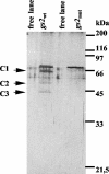
CHLAMY 1 is composed of different subunits. CHLAMY 1 was purified in parallel with either the gs2wt or gs2mut transcript as described for Fig. 1A. Eluted proteins were dialyzed, electrophoresed on an SDS-12.5% polyacrylamide gel along with a molecular weight standard, and visualized by silver staining. Two free lanes demonstrate background bands caused by the silver staining procedure.
FIG. 3.
FIG. 3.
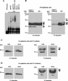
Protein-protein interaction of the C1 and C3 subunits and their interaction with the UG-containing gs2wt transcript. (A) Autoradiogram of supershift assays using the 32P-labeled gs2wt transcript. The first lane labeled with RNA demonstrates the mobility of the transcript (gs2wt) alone. For the binding reaction, the samples were incubated in the presence of poly(G) with a crude extract (CE lanes) from Chlamydomonas cells that were harvested at the beginning of the night (LD 14). In some cases, anti-C3 antibody (lane 3) and anti-C1/2 antibody (lane 4) were added to the binding reaction as described in Materials and Methods. In one case, anti-C1/2 and anti-C3 antibodies were added together to the binding reaction (lane 5). As a negative control, C3 (lane 6) and C1/2 (lane 7) preimmune sera (PIS) were added to the binding reaction. RNA-protein complexes were resolved in a nondenaturing 4% polyacrylamide gel containing 10% glycerol. (B) Coimmunoprecipitation assays were carried out with proteins from crude extracts of Chlamydomonas cells, which had been grown in continuous light. Precipitation was done according to the procedure described in Materials and Methods with polyclonal antibodies directed against the C3 subunit or with preimmune serum as control. Precipitated proteins were denatured, separated by SDS-9% PAGE, and subjected to a Western blot analysis with antibodies directed either against the C3 (C3 detection) or the C1 (C1 detection) subunit. Cross-reactions of immunoglobulin G (IgG) with the secondary antibody (see Materials and Methods) are indicated. (C and D) Coimmunoprecipitation assays were carried out with proteins from crude extracts of Chlamydomonas cells, which were grown under a LD cycle and harvested either at LD 6 or at LD 14. Coimmunoprecipitation was performed with the antibodies directed against either the C3 (C) or the C1 (D) subunit. Precipitated proteins were treated as described for panel B and examined in Western blot analysis for the presence of both subunits (C3 and C1 detection).
FIG. 4.
FIG. 4.
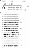
Domain architecture and amino acid composition of the protein encoded by the c1 cDNA. (A) Positions of the 5′ UTR, ORF, and the 3′ UTR are shown. It should be noted that the 5′ UTR may not be complete. The two arrows indicate a sequence part, which is not contained in our c1 cDNA clone, whose sequence can be found under GenBank accession number AY505473. Sequences of this N-terminal part have been obtained from the EST clone BI721496 (length, 579 bp), which has 381 bp overlapping with our cDNA clone. A BLAST search had been carried out for conserved domain architecture. Relevant domains are labeled. KH domains are known to be involved in RNA binding; the WW domain is a protein-protein interaction domain. G/P/Y indicates a region which is rich in Gly, Pro, and Tyr. The positions of the sequenced peptides (see Table 1) are indicated by gray (deriving from the C1 subunit) and black (deriving from the C2 subunit) boxes. Peptide 8* (from C2) overlaps in its sequence with peptide 16 (from C1). (B) The amino acid sequence of the c1 ORF is presented. The KH and WW domains have been compared to their standard domains (smart accession no. SM0322 for KH and SM0456 for WW). Identical amino acids have been highlighted in black, functionally similar amino acids in gray. The amino acids Gly, Pro, and Tyr, which frequently appear between the third KH and the WW domain, are underlined.
FIG. 5.
FIG. 5.
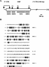
Domain architecture and amino acid composition of the protein encoded by the c3 cDNA. (A) Positions of the 5′ UTR, ORF, and the 3′ UTR are shown. It should be noted that the 5′ UTR may not be complete. The two arrows indicate a sequence part, which is not contained in our c3 cDNA clone, whose sequence can be found under GenBank accession number AY505474. Sequences of this N-terminal part have been obtained from the EST clone AV641734 (length, 506 bp), which has 168 bp overlapping with our cDNA clone. A BLAST search had been carried out for conserved domain architecture. Relevant domains are labeled. RRM domains are known to be involved in RNA binding. G and M indicate a region which is rich in Met and Gly. The positions of the sequenced peptides (see Table 1) are indicated by black boxes. (B) The amino acid sequence of the c3 ORF is presented. The RRM domains have been compared to their standard domain (smart accession no. SM0360). Identical amino acids have been highlighted in black, functionally similar amino acids in gray. The amino acids Met and Gly, which frequently appear between the second and third RRM domain, are indicated in bold (Met) or are underlined (Gly).
FIG. 6.
FIG. 6.
The amounts of the C1 and C3 subunits do not correlate with circadian binding activity of CHLAMY 1. Chlamydomonas cells were grown under a light-dark cycle (LD) or under continuous dim light (LL) and cells were harvested at the indicated time points. Proteins from a crude extract were denatured and 100 μg per lane was electrophoresed by SDS-9% PAGE. Western blot analysis was carried out by using the antibodies directed against either the C3 or the C1 subunit. To control for equal amounts of loaded proteins, another gel was run in parallel and the proteins were stained by Coomassie stain (data not shown).
FIG. 7.
FIG. 7.
Formation of multiprotein complexes bearing the C1 and C3 subunits during day and night phases. Cells were grown under a LD cycle and then put under LL. They were harvested in the middle of subjective day (LL 28) and at the beginning of subjective night (LL 38). Crude extracts were prepared according to the procedure in Materials and Methods. For each gradient, equal amounts of proteins were loaded on a linear 6 to 14% sucrose density gradient. The gradients were centrifuged at 74,100 × g for 16 h at 4°C. Fraction numbering starts with the low percentage (6%). In parallel, a gradient with proteins of known molecular masses was run. Aliquots of the fractions were electrophoresed by SDS-9% PAGE and analyzed in Western blots for the presence of the C3 (A) and C1 (B) subunits.
FIG. 8.
FIG. 8.
The C3 subunit shows significant homology to the rat CUG-BP2 (52 kDa) and belongs to the CELF family. The anti-C3 antibody of C. reinhardtii can recognize a 52-kDa protein in different rat brain tissues and in the retina. (A) The amino acid sequence of C3 was aligned with rat CUG-BP2 (NP_058893) by using the DNA Star program by Clustal W. Identical amino acids are marked in black, functionally similar ones in gray. The positions of the three RRM domains are indicated. (B) Rats were sacrificed at the beginning of the day (LD 2) and different brain tissues were taken. Crude extracts of rat brain tissues were prepared as described in Materials and Methods. Proteins were electrophoresed by SDS-9% PAGE and used for Western analysis with either preimmune serum (PIS) as control or anti-C3 antibody (C3).
FIG. 8.
FIG. 8.
The C3 subunit shows significant homology to the rat CUG-BP2 (52 kDa) and belongs to the CELF family. The anti-C3 antibody of C. reinhardtii can recognize a 52-kDa protein in different rat brain tissues and in the retina. (A) The amino acid sequence of C3 was aligned with rat CUG-BP2 (NP_058893) by using the DNA Star program by Clustal W. Identical amino acids are marked in black, functionally similar ones in gray. The positions of the three RRM domains are indicated. (B) Rats were sacrificed at the beginning of the day (LD 2) and different brain tissues were taken. Crude extracts of rat brain tissues were prepared as described in Materials and Methods. Proteins were electrophoresed by SDS-9% PAGE and used for Western analysis with either preimmune serum (PIS) as control or anti-C3 antibody (C3).
FIG. 9.
FIG. 9.
The rat C3 homologue is also present in protein complexes. Rats were sacrificed at the beginning of the day (LD 2). A crude extract of the cortex was prepared according to the procedure from Materials and Methods. Aliquots of the fractions were loaded on a linear 6 to 14% sucrose density gradient (for details, see the legend for Fig. 7). Aliquots of the fractions were electrophoresed by SDS-9% PAGE and analyzed in Western blots with anti-C3 antibody for the presence of the 52-kDa rat C3 homologue. Due to slightly smaller tubes (see Materials and Methods), peaks of the standard proteins do not migrate to the same positions as the ones from the sucrose gradients of Fig. 7.

Similar articles

Cited by

References

    1. Adams, P. D., S. Seeholzer, and M. Ohn. 2002. Identification of associated proteins by coimmunoprecipitation. Protein-protein interaction: a molecular cloning manual, p. 59-74. Cold Spring Harbor Laboratory Press, Cold Spring Harbor, N.Y.
    1. Adinolfi, S., A. Ramos, S. R. Martin, P. F. Dal, P. Pucci, B. Bardoni, J. L. Mandel, and A. Pastore. 2003. The N-terminus of the fragile X mental retardation protein contains a novel domain involved in dimerization and RNA binding. Biochemistry 42:10437-10444. - PubMed
    1. Allada, R., P. Emery, J. S. Takahashi, and M. Rosbash. 2001. Stopping time: the genetics of fly and mouse circadian clocks. Annu. Rev. Neurosci. 24:1091-1119. - PubMed
    1. Buratti, E., and F. B. Baralle. 2001. Characterization and functional implications of the RNA binding properties of nuclear factor TDP-43, a novel splicing regulator of CFTR exon 9. J. Biol. Chem. 276:36337-36343. - PubMed
    1. Burd, C. G., and G. Dreyfuss. 1994. Conserved structures and diversity of functions of RNA-binding proteins. Science 265:615-621. - PubMed

Publication types

MeSH terms

LinkOut - more resources