. 2004 Jun 9;24(23):5292-300.
doi: 10.1523/JNEUROSCI.0195-04.2004.

Microdomains for dopamine volume neurotransmission in primate prefrontal cortex

Affiliations

Microdomains for dopamine volume neurotransmission in primate prefrontal cortex

Constantinos D Paspalas et al. J Neurosci. .

Abstract

The explicit yet enigmatic involvement of dopamine in cortical physiology is in part volumetric (beyond the synapse), as is apparently the action of neuroleptics targeting dopamine receptors. The notion that nonsynaptic neuronal membranes would translate extracellular dopamine into receptor-specific spatiotemporal downstream signaling, similar to the chemical synapse, is intriguing. Here, we report that dopamine D5 (but not D1 or D2) receptors in the perisomatic plasma membrane of prefrontal cortical neurons form discrete and exclusively extrasynaptic microdomains with inositol 1,4,5-trisphosphate-gated calcium stores of subsurface cisterns and mitochondria. These findings introduce a novel dopaminoceptive substratum in the brain and a unique D5 receptor-specific signaling paradigm.


Figures

Figure 1.
Figure 1.
A, B, Schematic representation of the relative topography of the SSC (A; arrowheads in frame and enlarged schema) and electron micrograph depicting oblique plane of section through the plasmalemma-SSC interface (B). As seen in B, SSCs typically consist of single or multiple flattened cisterns (electron opaque zone subjacent to the membrane; arrowheads), confluent through anastomoses with smooth reticular endomembranes (SER) and continuous with the rough endoplasmic reticulum (arrows). Therefore, an SSC derives from the reticular network of which is considered spatial and potentially functional modification. In the CNS and peripheral nervous system, SSCs are neuron specific and localized exclusively in perikarya and the most proximal portions of their processes (Rosenbluth, 1962; Henkart et al., 1976; Peters et al., 1991). Likewise, SSCs occur in perikarya and large-caliber proximal dendrites of corticalneurons (Rosenbluth, 1962; Buschmann, 1979; for review, see Peters et al., 1991) and are unique to the axon initial segment of principal cells (Palay et al., 1968; Benedeczky et al., 1994). Double arrowheads in B point to an axo-spinous synapse (ax-sp) next to the SSC. mit, Mitochondrion. Scale bar, 100 nm.
Figure 5.
Figure 5.
Subcellular distribution of InsP3Rs (arrows) in the neuropil and the perisomatic region (see supplemental Fig. 1, available at www.jneurosci.org) and coexpression with D5Rs (arrowheads). A, The spine apparatus (asterisk) of a dendritic spine (sp) displays InsP3R immunoreactivity with a predilection for saccules facing the postsynaptic density of an apposed synaptic varicosity; the obliquely sectioned synaptic cleft is not visible. B, Inset, InsP3R immunoparticles decorate slender SER tubules in dendrites (den). In the perisomatic region, InsP3Rs were rarely visualized on the cis-face of Golgi complexes (C), whereas the main body of immunoreactivity appears on the cytoplasmic face of reticular endomembranes (D; compare with the large D5R immunoparticles of the cytoplasmic pool). Note the loose association of a cisternal profile (D, double arrowheads) with an axo-somatic symmetric synapse. Elaborate cisternal appositions have been described to accompany symmetric GABA ergic synapses on the axon initial segment of hippocampal pyramids and to sequester free Ca2+ (Benedeczky et al., 1994). ax, Axon. Scale bars, 200 nm (applies also to B, inset).
Figure 6.
Figure 6.
Coexpression of D5Rs and InsP3Rs in microdomains. In A, D5Rs were visualized with immunoperoxidase precipitates. Despite the diffuse nature of the immunomarker, two plasmalemma-SSC complexes (framed) and the subjacent cytoplasm demonstrate increased labeling density compared with uncomplexed membranes or neighboring cellular profiles. InsP3Rs were simultaneously visualized with nanogold at high magnification (inset, arrows). To avoid artifactual contrasting of membranes, this material was processed with minimal lead counterstaining. For illustration purposes, image contrast in A and inset has been enhanced digitally (Photoshop 7.0; Adobe Systems, San Jose, CA). When detected with nanogold probes (arrowheads), D5Rs were visualized between the plasmalemma and juxtaposed cisternal endomembranes (C, inset, G), and predominantly at the periphery of plasmalemma-SSC complexes (D, E). The InsP3R-immunoreactive profiles (arrows) included the lateral vacuolar expansions of SSCs (B--E) and their middle portion (A, F,G, inset, H). Note in C the reversal of the immunolabeling sequence for D5R (small particles; main panel and inset) and InsP3R (large particles); double arrowheads point to the plasmalemma-SSC interface. H illustrates a rare case of somato-somatic membrane juxtaposition furnished with two SSCs (white arrowheads); one SSC is confluent with the reticular network (double arrowheads). Notice that the plasmalemma-SSC complex, which is immunoreactive for both D5Rs and InsP3Rs, also coincides spatially with the widening of the perineuronal space (asterisks), perhaps to allow access via extracellular diffusion. White arrow in B points to a dense-cored vesicle within a DAergic-like axon (ax) deployed en passant. Scale bars: A, 500 nm; A inset, B, D--G, 200 nm; G inset, 20 nm; C, 100 nm; C inset, 25 nm.
Figure 2.
Figure 2.
Differential distribution of DAR subtypes in the perisomatic region. Dashed lines and arrowheads outline uncomplexed plasma membrane and plasmalemma-SSC complexes, respectively. A, D1Rs are exclusively associated with the Golgi apparatus with a marked predilection for certain Golgi complexes. B, C, Similarly, the D2Rs comprise an intracellular pool, often in association with endosomes (B, arrows) but also a plasmalemmal component on uncomplexed membranes (C, arrows). D--G, In contrast, D5Rs form distinct cytoplasmic clusters, including clathrin-coated vesicles (D, arrows) subjacent to plasmalemma-SSC complexes; note in D a nearby immunonegative complex. D5Rs are additionally expressed on membranes flanking SSCs (F, G), as contrasted with uncomplexed and synaptic (D, double arrowheads) membranes. E and F depict consecutive sections. Arrows in D, inset, point to the plasmalemma and to individual cisternal profiles; a particle aggregate (arrowhead) is likely displaced from the somatic membrane, for apposed cellular profiles were rarely immunoreactive. ax, Axon; den, dendrite; lys, lysosome; mvb, multivesicular body. Scale bars: A, B, D--G, 400 nm; C, D, inset, 200 nm.
Figure 3.
Figure 3.
D5R expression in the perisomatic region. All panels depict nonpermeabilized material, hence the lack of immunolabeling in the plasmalemma-SSC interface (compare with Fig. 4). A-E, In perikarya (A, B, E) and primary dendrites (den; C, D), D5Rs are selectively clustered subjacent to plasmalemma-SSC complexes (A, B, arrowheads) or disposed on flanking membranes. Uncomplexed and synaptic (A, B, double arrowheads) membranes are devoid of immunoreactivity. Sections shown in C and D belong to a series and are 280 nm apart. Notice how the mitochondrion (mit) in C appears in D directly apposed to the SSC. In both micrographs, glial leaflets (asterisks) separate the plasmalemma-SSC complex from an apposed varicosity (ax) forming an asymmetric axo-spinous synapse (double arrowheads). In C-E, arrowheads point to periodic cross-bridges in the plasmalemma-SSC interface of similar size and periodicity to the tetrameric protrusions described in Purkinje SER membranes with freeze-fracture/replication (Kanaseki et al., 1998). It has been suggested that these electron-dense protrusions, which also appear in plasmalemma-SSC complexes of hippocampal pyramids (Benedeczky et al., 1994), may correspond to the fourfold symmetry of the tetrameric InsP3R (Kanaseki et al., 1998; Jiang et al., 2002). Scale bars: A, 300 nm; B-E, 200 nm.
Figure 4.
Figure 4.
D5R immunoreactivity visualized with single particles after membrane permeabilization. A-D, Arrows point to the receptor cytoplasmic pool, and arrowheads point to the plasmalemmal component on membranes overlying (A--C) or flanking (D) SSCs. Notice how this architecture confines a 10-15 nm wide cytosolic microcompartment (double arrowheads) between the plasma membrane and the cisternal outer limiting membrane. White arrowheads point to an oblique section of the plasmalemma (and the SSC); the cytosolic microcompartment is not visible. Scale bars, 200 nm.
Figure 7.
Figure 7.
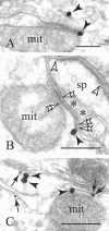
The D5R microdomains establish intimate associations with mitochondria (mit). A, B, D5Rs (black arrowheads) are localized at the edges of the microdomain and on a neuropil profile (A). A dendritic spine (sp) receiving an asymmetric synapse (between arrowheads in B) is apposed to the microdomain with intervening glial processes (asterisks); double arrowheads point to cross-bridges (Fig. 3 legend) in the plasmalemma-SSC and SSC-mitochondrion interface. C depicts InsP3R expression (two attached immunoparticles; arrows) within the minute cleft separating the cisternal from the outer mitochondrial membrane. Scale bars, 200 nm.

Similar articles

Cited by

References

    1. Agnati LF, Bjelke B, Fuxe K (1995a) Volume versus wiring transmission in the brain: a new theoretical frame for neuropsychopharmacology. Med Res Rev 15: 33-45. - PubMed
    1. Agnati LF, Zoli M, Strömberg I, Fuxe K (1995b) Intercellular communication in the brain: wiring versus volume transmission. Neuroscience 69: 711-726. - PubMed
    1. Agnati LF, Fuxe K, Nicholson C, Syková E (2000) Progress in brain research, Vol 125. Volume transmission revisited. Amsterdam: Elsevier.
    1. Alonso-Nanclares L, Minelli A, Melone M, Edwards RH, DeFelipe J, Conti F (2004) Perisomatic glutamatergic axon terminals: a novel feature of cortical synaptology revealed by vesicular glutamate transporter 1 immunostaining. Neuroscience 123: 547-556. - PubMed
    1. Ariano MA, Wang J, Noblett KL, Larson ER, Sibley DR (1997) Cellular distribution of the rat D1B receptor in central nervous system using anti-receptor antisera. Brain Res 746: 141-150. - PubMed

Publication types

MeSH terms

LinkOut - more resources