. 2004 Jun 9;24(23):5370-80.
doi: 10.1523/JNEUROSCI.1219-04.2004.

Identification of sympathetic premotor neurons in medullary raphe regions mediating fever and other thermoregulatory functions

Affiliations

Identification of sympathetic premotor neurons in medullary raphe regions mediating fever and other thermoregulatory functions

Kazuhiro Nakamura et al. J Neurosci. .

Abstract

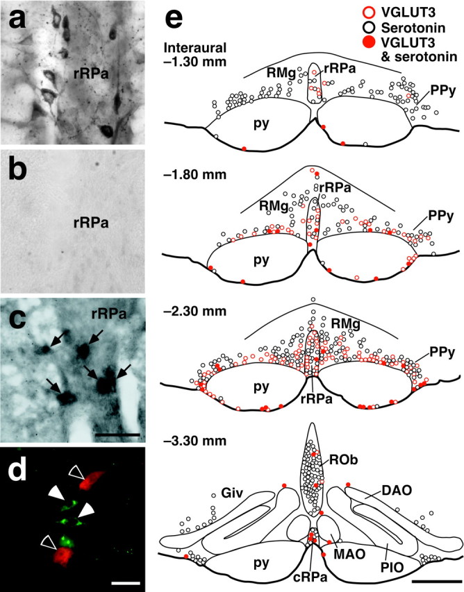
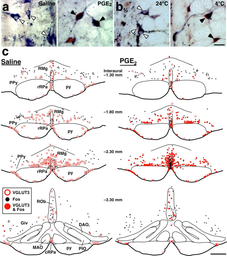
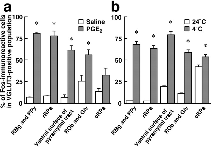
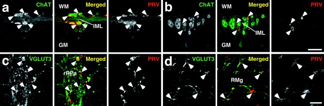
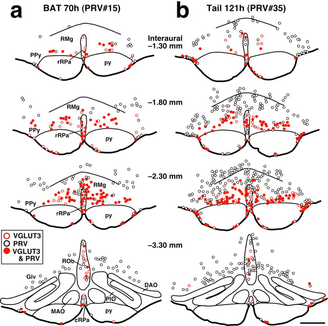
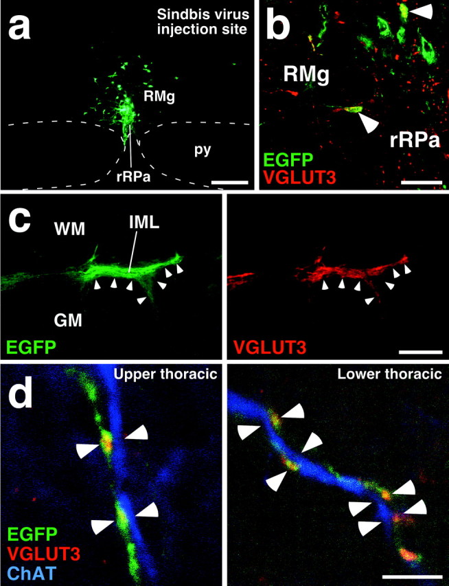
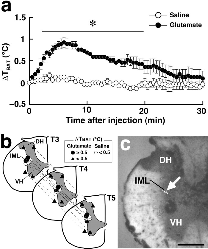
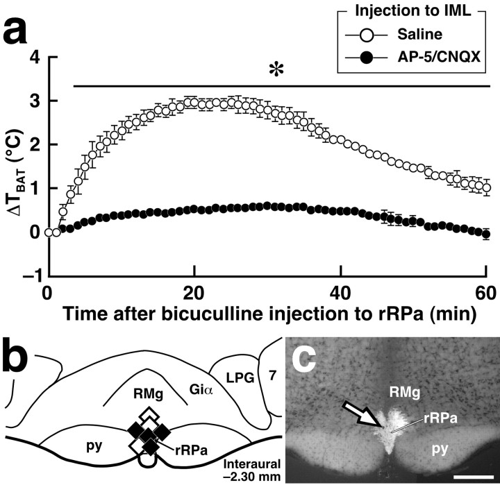
Sympathetic premotor neurons directly control sympathetic preganglionic neurons (SPNs) in the intermediolateral cell column (IML) of the thoracic spinal cord, and many of these premotor neurons are localized in the medulla oblongata. The rostral ventrolateral medulla contains premotor neurons controlling the cardiovascular conditions, whereas rostral medullary raphe regions are a candidate source of sympathetic premotor neurons for thermoregulatory functions. Here, we show that these medullary raphe regions contain putative glutamatergic neurons and that these neurons directly control thermoregulatory SPNs. Neurons expressing vesicular glutamate transporter 3 (VGLUT3) were distributed in the rat medullary raphe regions, including the raphe magnus and rostral raphe pallidus nuclei, and mostly lacked serotonin immunoreactivity. These VGLUT3-positive neurons expressed Fos in response to cold exposure or to central administration of prostaglandin E2, a pyrogenic mediator. Transneuronal retrograde labeling after inoculation of pseudorabies virus into the interscapular brown adipose tissue (BAT) or the tail indicated that those VGLUT3-expressing medullary raphe neurons innervated these thermoregulatory effector organs multisynaptically through SPNs of specific thoracic segments, and microinjection of glutamate into the IML of the BAT-controlling segments produced BAT thermogenesis. An anterograde tracing study further showed a direct projection of those VGLUT3-expressing medullary raphe neurons to the dendrites of SPNs. Furthermore, intra-IML application of glutamate receptor antagonists blocked BAT thermogenesis triggered by disinhibition of the medullary raphe regions. The present results suggest that VGLUT3-expressing neurons in the medullary raphe regions constitute excitatory neurons that could be categorized as a novel group of sympathetic premotor neurons for thermoregulatory functions, including fever.


Figures

Figure 1.
Figure 1.
Expression of VGLUT3 in medullary raphe neurons. a, Immunoperoxidase staining for VGLUT3 in medullary raphe regions. VGLUT3 immunoreactivity was localized in neuronal cell bodies as well as fibers and terminals. b, No immunoreactivity was observed after preincubation of the anti-VGLUT3 antibody with the antigenic peptide. c, In situ hybridization for VGLUT3 mRNA in medullary raphe regions. Hybridization signals for VGLUT3 mRNA were exhibited by medullary raphe neurons (arrows). d, Double immunofluorescence labeling for VGLUT3 (green) and serotonin (red) in medullary raphe regions. VGLUT3-immunoreactive neurons (filled arrowheads) and serotonin-immunoreactive neurons (open arrowheads) were clearly visualized. e, Brain maps showing the distribution of VGLUT3-immunoreactive neurons and serotonin-immunoreactive ones. Immunoreactive cell bodies in a 20-μm-thick frontal section of the corresponding rostrocaudal position were plotted on a drawing. DAO, Dorsal accessory olivary nucleus; MAO, medial accessory olivary nucleus; PIO, principal inferior olivary nucleus; py, pyramidal tract. Scale bars: a-c (in c), 50 μm; d, 30 μm; e, 500 μm.
Figure 2.
Figure 2.
Fos expression in VGLUT3-immunoreactive medullary raphe neurons in response to central PGE2 application and cold exposure. a, b, Double immunoperoxidase staining for VGLUT3 (blue-black) and Fos (brown) in the rRPa and RMg after intracerebroventricular injection of saline or PGE2 (a) and after exposure of rats to environmental temperature of 24 or 4°C (b). The open and filled arrowheads indicate VGLUT3-immunoreactive neuronal cell bodies that are negative and positive for Fos immunoreactivity, respectively. c, Brain maps showing the distribution of VGLUT3-immunoreactive neurons and Fos-immunoreactive cells. Immunoreactive cells in a 20-μm-thick frontal section of the corresponding rostrocaudal position were plotted on a drawing. Scale bars: a, b (in b), 20 μm; c, 500 μm.
Figure 3.
Figure 3.
Quantitative analysis of Fos expression in VGLUT3-immunoreactive medullary raphe neurons in response to intracerebroventricular injection of saline (open bars; n = 3) or PGE2 (shaded bars; n = 4) (a) and to exposure of rats to environmental temperature of 24°C (open bars; n = 3) or 4°C (shaded bars; n = 3) (b). The number of immunoreactive cells was counted in every six 20-μm-thick frontal sections of the medulla oblongata (see Fig. 2). Each bar represents the mean ± SEM of the group. Asterisks indicate statistically significant differences between the PGE2- and saline-injected groups or between the 4°C- and 24°C-exposed groups (p < 0.05).
Figure 4.
Figure 4.
Retrograde transneuronal labeling with PRV inoculated into the interscapular BAT or the tail. a, b, Double immunofluorescence staining for ChAT (green) and PRV (red) in horizontal spinal cord sections of the T3 segment at 47 hr after inoculation into the BAT (a) and of the T12 segment at 98 hr after inoculation into the tail (b). Arrowheads indicate PRV-infected ChAT-immunoreactive neurons clustering in the IML. c, d, Double immunofluorescence staining for VGLUT3 (green) and PRV (red) in rostral medullary raphe regions in frontal sections at 69 hr after inoculation into the BAT (c) and at 121 hr after inoculation into the tail (d). The arrowheads indicate PRV-infected VGLUT3-immunoreactive neurons. GM, Gray matter; WM, white matter. Scale bars: a, b (in b), 100 μm; c, d (in d), 50 μm.
Figure 5.
Figure 5.
Brain maps showing the distribution of VGLUT3-immunoreactive neurons and PRV-immunoreactive ones at 70 hr after inoculation into the interscapular BAT (a) and at 121 hr after inoculation into the tail (b). Immunoreactive cell bodies in a 20-μm-thick frontal section of the corresponding rostrocaudal position were plotted on a drawing. PRV inoculation was made on the right side. Scale bar (in b), 500 μm.
Figure 6.
Figure 6.
Direct projection of VGLUT3-immunoreactive medullary raphe neurons onto SPNs. a, Injection of recombinant Sindbis virus into the rRPa and RMg at the rostrocaudal level of interaural -2.30 mm. Infected cells expressed a membrane-targeted form of EGFP. b, Infection of VGLUT3-immunoreactive neurons with the Sindbis virus. There were neurons exhibiting both VGLUT3 immunoreactivity (red) and EGFP fluorescence (green) in the injection site (arrowheads). c, Double fluorescence microscopy for EGFP fluorescence and VGLUT3 immunoreactivity in a horizontal spinal cord section of the T3 segment. The right and left photomicrographs were taken at the same field under different excitations. EGFP-positive axon fibers and terminals were densely localized in the IML, and their distribution well overlapped that of VGLUT3-immunoreactive terminals (arrowheads). d, Confocal laser-scanning microscopy of IML sections triple immunofluorescence-stained for EGFP (green), VGLUT3 (red), and ChAT (blue). Close apposition of axon swellings double-labeled with EGFP and VGLUT3 immunoreactivities to ChAT-immunoreactive dendrites (arrowheads) was found in both upper (T1-T4) and lower (T10-T13) thoracic segments. Scale bars: a, 300 μm; b, 50 μm; c, 200 μm; d, 5 μm.
Figure 7.
Figure 7.
BAT thermogenesis induced by glutamate microinjection into the IML. a, Changes in temperature of the interscapular BAT (TBAT) after saline or glutamate microinjections into the IML at three sites in segments T3, T4, and T5 (b, circles). TBAT was monitored at intervals of 30 sec. Each value represents the mean ± SEM of three rats per group. The changes in TBAT were significantly different between the glutamate- and saline-injected groups during the time period denoted by a horizontal bar with an asterisk (p < 0.05). b, Composite drawing of the effect of glutamate or saline microinjections into the thoracic spinal cord on TBAT. Each symbol group tied with dotted lines corresponds to sites of microinjections made in one experiment and represents corresponding changes in TBAT 30 min after the microinjections. c, A representative view of a site of intra-IML microinjection. The injection site is clearly identified as a small cluster of fluorescent beads (arrow) in the transverse section counterstained with toluidine blue. DH, Dorsal horn; VH, ventral horn. Scale bar, 500 μm.
Figure 8.
Figure 8.
Microinjections of glutamate receptor antagonists into the IML block BAT thermogenesis triggered by bicuculline-induced disinhibition of rostral medullary raphe neurons. a, Changes in TBAT after bicuculline injection into the rRPa in rats microinjected with a mixture of AP-5 and CNQX (n = 4) or saline (n = 3) into the IML over the T2-T6 segments. TBAT was monitored at intervals of 1 min. Each value represents the mean ± SEM of the group. The changes in TBAT were significantly different between the AP-5/CNQX- and saline-pretreated groups during the time period denoted by a horizontal bar with an asterisk (p < 0.05). b, The position of the bicuculline (filled diamonds) and saline (open diamonds) injections made in a. c, A representative view of a site of intra-rRPa injection at the level of interaural -2.30 mm. The injection site is clearly identified as a cluster of fluorescent beads (arrow) in the section counterstained with toluidine blue. 7, Facial nucleus; Giα, alpha part of the gigantocellular reticular nucleus; LPG, lateral paragigantocellular nucleus. Scale bar, 500 μm.

Similar articles

Cited by

References

    1. Aicher SA, Hahn B-I, Milner TA (2000) N-methyl-d-aspartate-type glutamate receptors are found in post-synaptic targets of adrenergic terminals in the thoracic spinal cord. Brain Res 856: 1-11. - PubMed
    1. Bamshad M, Song CK, Bartness TJ (1999) CNS origins of the sympathetic nervous system outflow to brown adipose tissue. Am J Physiol 276: R1569-R1578. - PubMed
    1. Banet M, Hensel H, Liebermann H (1978) The central control of shivering and non-shivering thermogenesis in the rat. J Physiol (Lond) 283: 569-584. - PMC - PubMed
    1. Blessing WW, Nalivaiko E (2001) Raphe magnus/pallidus neurons regulate tail but not mesenteric arterial blood flow in rats. Neuroscience 105: 923-929. - PubMed
    1. Blessing WW, Yu YH, Nalivaiko E (1999) Raphe pallidus and parapyramidal neurons regulate ear pinna vascular conductance in the rabbit. Neurosci Lett 270: 33-36. - PubMed

Publication types

MeSH terms