

Somatotopy and attentional modulation of the human parietal and opercular regions
- PMID: 15190112
- PMCID: PMC6729293
- DOI: 10.1523/JNEUROSCI.4030-03.2004
Somatotopy and attentional modulation of the human parietal and opercular regions
Abstract
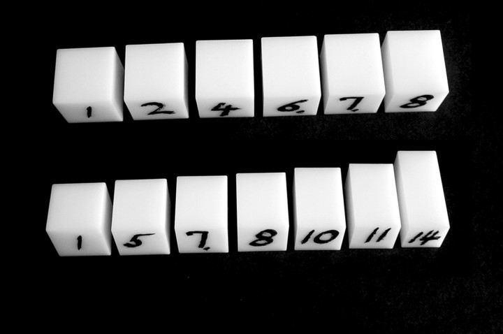
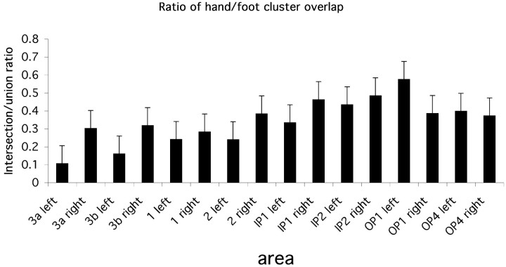
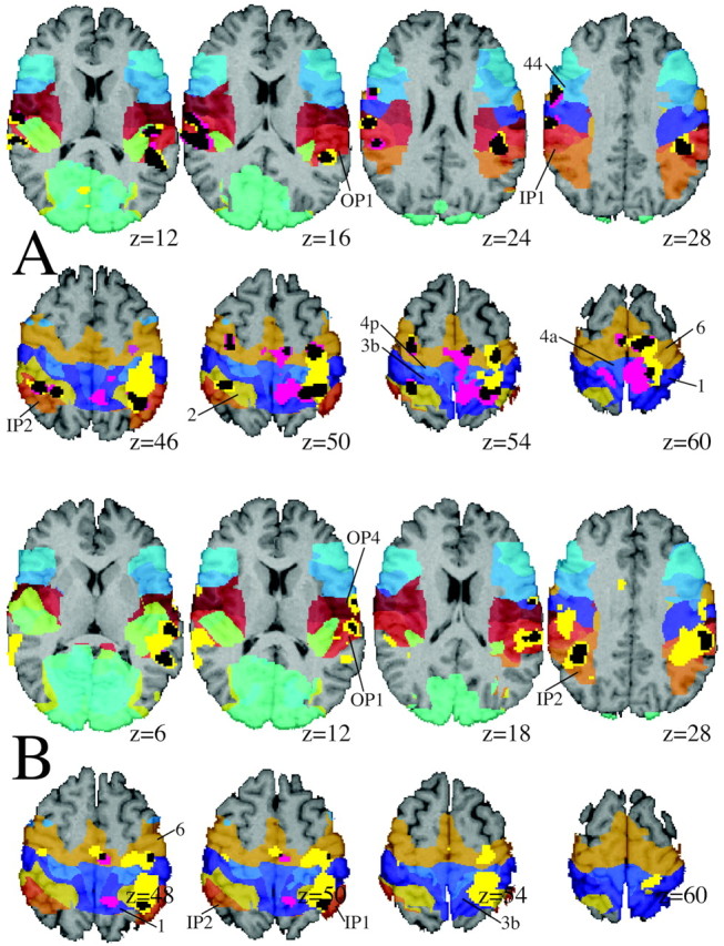
The somatotopical organization of the postcentral gyrus is well known, but less is known about the somatotopical organization of area 2, the somatosensory association areas in the postparietal cortex, and the parietal operculum. The extent to which these areas are modulated by attention is also poorly understood. For these reasons, we measured the BOLD signal when rectangular parallelepipeds of varying shape were presented to the immobile right hand or right foot of 10 subjects either discriminating these or just being stimulated. Activation areas in each subject were mapped against cytoarchitectural probability maps of area 2, IP1, and IP2 along the intraparietal sulcus and the parietal opercular areas OP1-OP4. In area 2, the somatotopical representation of the hand and foot were distinctly separate, whereas there was considerable overlap in IP1 and no clear evidence of separate representations in OP1, OP4, and IP2. The overlap of hand and foot representations increased in the following order: area 3a, 3b, 1, 2, IP1, OP4, IP2, and OP1. There were significant foot representations but no hand representations in right (ipsilateral) areas 3a, 3b, and 1. Shape discrimination using the foot as opposed to stimulation enhanced the signal in OP4 bilaterally, whereas discrimination with the hand enhanced the signal bilaterally in area 2, IP1, and IP2. These results indicate that somatosensory areas in humans are arranged from strong somatotopy into no somatotopy in the following order: 3a, 3b, 1, 2, IP1, OP4, IP2, and OP1. Higher order areas such as IP1, IP2, and OP4 showed task-related attentional enhancement.
Figures


Similar articles
-
Anatomical and functional properties of the foot and leg representation in areas 3b, 1 and 2 of primary somatosensory cortex in humans: A 7T fMRI study.Neuroimage. 2017 Oct 1;159:473-487. doi: 10.1016/j.neuroimage.2017.06.021. Epub 2017 Jun 17. Neuroimage. 2017. PMID: 28629975
-
Cortical network for vibrotactile attention: a fMRI study.Hum Brain Mapp. 2008 Feb;29(2):207-21. doi: 10.1002/hbm.20384. Hum Brain Mapp. 2008. PMID: 17390318 Free PMC article.
-
The organization and connections of somatosensory cortex in marmosets.J Neurosci. 1990 Mar;10(3):952-74. doi: 10.1523/JNEUROSCI.10-03-00952.1990. J Neurosci. 1990. PMID: 2108231 Free PMC article.
-
The functional organization of somatosensory cortex in primates.Ann Anat. 1993 Dec;175(6):509-18. doi: 10.1016/s0940-9602(11)80212-8. Ann Anat. 1993. PMID: 8297039 Review.
-
Parietofrontal circuits for action and space perception in the macaque monkey.Neuroimage. 2001 Jul;14(1 Pt 2):S27-32. doi: 10.1006/nimg.2001.0835. Neuroimage. 2001. PMID: 11373129 Review.
Cited by
-
Somatotopy in the Human Somatosensory System.Front Hum Neurosci. 2018 Jun 12;12:235. doi: 10.3389/fnhum.2018.00235. eCollection 2018. Front Hum Neurosci. 2018. PMID: 29950980 Free PMC article.
-
Human MST but not MT responds to tactile stimulation.J Neurosci. 2007 Aug 1;27(31):8261-7. doi: 10.1523/JNEUROSCI.0754-07.2007. J Neurosci. 2007. PMID: 17670972 Free PMC article.
-
Identifying human parieto-insular vestibular cortex using fMRI and cytoarchitectonic mapping.Hum Brain Mapp. 2006 Jul;27(7):611-21. doi: 10.1002/hbm.20205. Hum Brain Mapp. 2006. PMID: 16281284 Free PMC article.
-
The sensory cortical representation of the human penis: revisiting somatotopy in the male homunculus.J Neurosci. 2005 Jun 22;25(25):5984-7. doi: 10.1523/JNEUROSCI.0712-05.2005. J Neurosci. 2005. PMID: 15976087 Free PMC article.
-
The cortical and cerebellar representation of the lumbar spine.Hum Brain Mapp. 2014 Aug;35(8):3962-71. doi: 10.1002/hbm.22451. Epub 2014 Jan 24. Hum Brain Mapp. 2014. PMID: 24464423 Free PMC article.
References
-
- Bodegard A, Geyer S, Naito E, Zilles K, Roland PE (2000) Somatosensory areas in man activated by moving stimuli: cytoarchitectonic mapping and PET. NeuroReport 11: 187-191. - PubMed
-
- Bodegard A, Geyer S, Grefkes C, Zilles K, Roland PE (2001) Hierarchical processing of tactile shape in the human brain. Neuron 31: 317-328. - PubMed
-
- Boussaoud D, Desimone R, Ungerleider LG (1991) Visual topography of area TEO in the macaque. J Comp Neurol 306: 554-575. - PubMed
-
- Burton DB, Chelune GJ, Naugle RI, Bleasel A (1996) Neurocognitive studies in patients with supplementary sensorimotor area lesions. Adv Neurol 70: 249-261. - PubMed
-
- Burton H, Videen TO, Raichle ME (1993) Tactile-vibration-activated foci in insular and parietal-opercular cortex studied with positron emission tomography: mapping the second somatosensory area in humans. Somatosens Mot Res 10: 297-308. - PubMed
Publication types
MeSH terms
LinkOut - more resources
Full Text Sources