. 2004 Jun 9;24(23):5427-38.
doi: 10.1523/JNEUROSCI.4449-03.2004.

Using a hybrid neural system to reveal regulation of neuronal network activity by an intrinsic current

Affiliations

Using a hybrid neural system to reveal regulation of neuronal network activity by an intrinsic current

Michael Sorensen et al. J Neurosci. .

Abstract

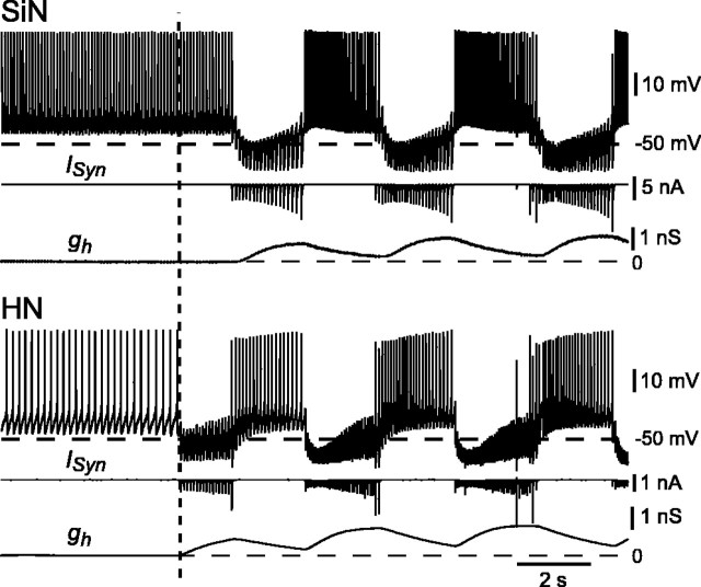
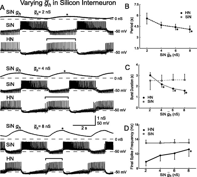
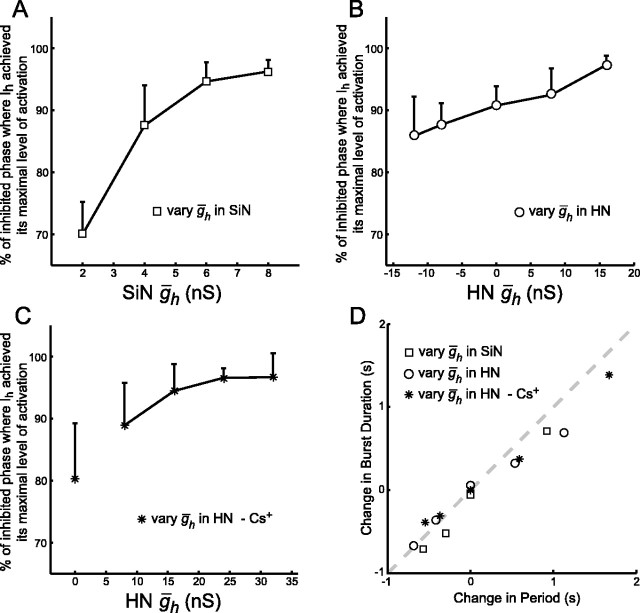
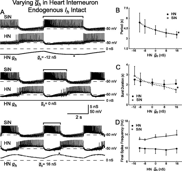
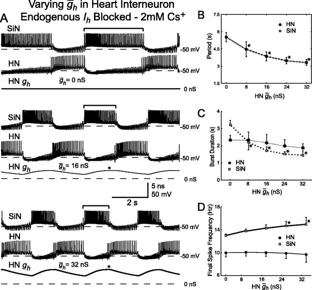
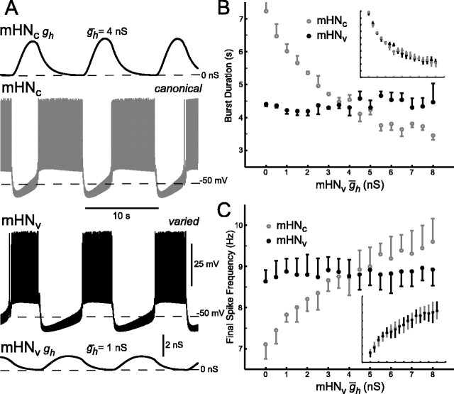
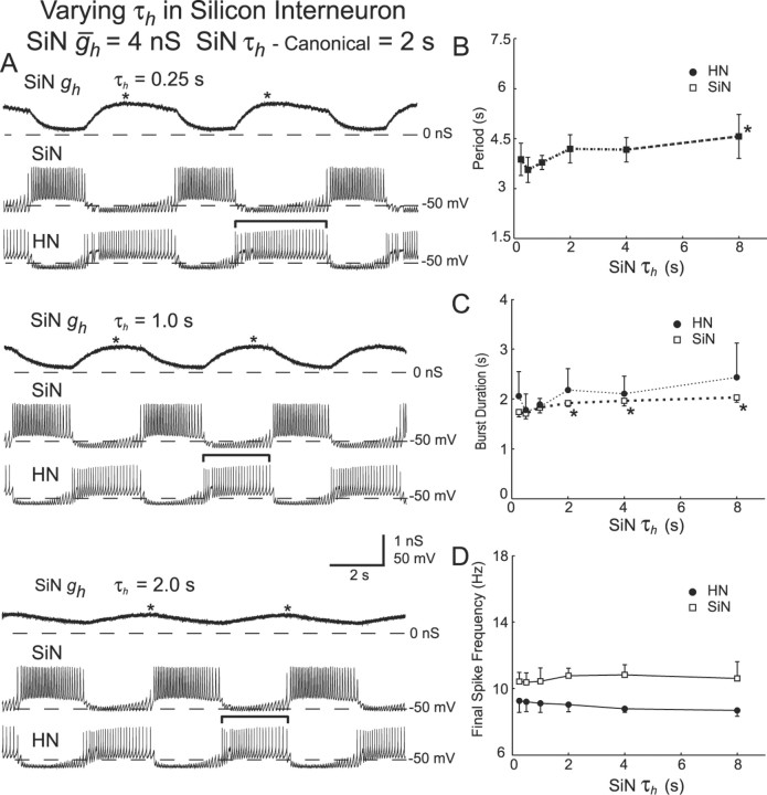
The generation of rhythmic patterns by neuronal networks is a complex phenomenon, relying on the interaction of numerous intrinsic and synaptic currents, as well as modulatory agents. To investigate the functional contribution of an individual ionic current to rhythmic pattern generation in a network, we constructed a hybrid system composed of a silicon model neuron and a heart interneuron from the heartbeat timing network of the medicinal leech. When the model neuron and a heart interneuron are connected by inhibitory synapses, they produce rhythmic activity similar to that observed in the heartbeat network. We focused our studies on investigating the functional role of the hyperpolarization-activated inward current (I(h)) on the rhythmic bursts produced by the network. By introducing changes in both the model and the heart interneuron, we showed that I(h) determines both the period of rhythmic bursts and the balance of activity between the two sides of the network, because the amount and the activation/deactivation time constant of I(h) determines the length of time that a neuron spends in the inhibited phase of its burst cycle. Moreover, we demonstrated that the model neuron is an effective replacement for a heart interneuron and that changes made in the model can accurately mimic similar changes made in the living system. Finally, we used a previously developed mathematical model (Hill et al. 2001) of two mutually inhibitory interneurons to corroborate these findings. Our results demonstrated that this hybrid system technique is advantageous for investigating neuronal properties that are inaccessible with traditional techniques.


Figures

Figure 1.
Figure 1.
Design and activity of hybrid half-center oscillator. A, Heart interneurons (HN), in an isolated ganglion preparation, were pharmacologically isolated with bicuculline. Voltage/current-clamp amplifiers were used for recording and current injection into heart interneurons and the silicon neuron (SiN). A single sharp microelectrode in discontinuous current-clamp mode was used for voltage recording and current injection into an isolated heart interneuron. Voltage recording and current injection into the silicon neuron were provided by direct connection to amplifier headstages (two-electrode current-clamp mode). Dynamic clamp provided real-time control signals to create artificial synapses between a heart interneuron and the silicon neuron. B, Activity of a living heart interneuron half-center oscillator in an isolated ganglion 3 (heart interneurons indexed by body side and ganglion number). C, Activity of a hybrid half-center oscillator.
Figure 2.
Figure 2.
Steady-state activation and inactivation curves for the silicon neuron. The circles represent data from voltage clamp. Solid lines denote best fit of data to the activation/inactivation equations of Hill et al. (2001). The dashed lines show steady-state activation and inactivation curves for the currents of the canonical mathematical heart interneuron model. A, Fast sodium current (INa). B, Inactivating potassium current (IK1). Data points from -75 to -45 mV are not shown because of large conductance/low current artifact. C, Slow, non-inactivating potassium current (IK2). D, Slowly inactivating low-threshold calcium current (ICaS). E, Persistent sodium current (IP). F, Hyperpolarization-activated inward current (Ih).
Figure 3.
Figure 3.
Activity of the silicon neuron depends on leak current parameters. A, Different types of activity, tonic spiking, bursting, and silence are expressed depending on the values of the leak reversal potential (Eleak) and the leak conductance (gleak). The regions of silence and tonic spiking are separated by a region of intrinsic bursting. A small region, indicated in black (multistability), where bursting coexisted with tonic spiking was also observed. Inset, Similar activity map for the mathematical model of a heart interneuron (Cymbalyuk et al., 2002a). B, Typical activity of the silicon neuron when firing tonically. C, Typical activity of the silicon neuron when bursting intrinsically. Leak parameter values for the activity in B and C are indicated on the map in A.
Figure 4.
Figure 4.
Dynamic-clamp-mediated inhibitory synapses of sufficient strength transform tonic firing into rhythmic antiphasic bursting in a hybrid half-center oscillator. A, Both the silicon neuron (SiN) and the pharmacologically isolated heart interneuron (HN) fire tonically in the absence of artificial synapses (ḡSyn = 0 nS). B, Weak artificial synapses (small ḡSyn) result in sporadic lapses in activity but not rhythmic bursting. C, Strong artificial synapses (large ḡSyn) result in stable alternating rhythmic bursting. The dashed line in A-C indicates -50 mV. D, Results of four experiments in which ḡSyn was varied between 0 and 625 nS. Different symbols represent different experiments. The period of rhythmic oscillations settle to a value that remains relatively unchanged with additional variations of ḡSyn. The error bars indicate the SD of the period over n ≥ 8 cycles for each trial of the four individual experiments.
Figure 5.
Figure 5.
Strong artificial inhibitory synapses cause an immediate transition from tonic firing to rhythmic bursting in a hybrid half-center oscillator. Membrane potential, synaptic current (ISyn), and h-current conductance (gh) are shown for the silicon neuron (SiN) and the heart interneuron (HN). The vertical dashed line indicates the time at which the dynamic clamp was activated to enable the mutual inhibitory synapses. When one of the neurons is inhibited, its Ih activates, driving it back toward the firing threshold. Once it begins to fire, the other neuron is inhibited, its Ih activates, driving it back toward firing threshold so that it inhibits the first neuron, and the cycle repeats.
Figure 6.
Figure 6.
Variation of ḡh in the silicon neuron (SiN ḡh) of a hybrid half-center oscillator. A, Typical activity at three different values of SiN ḡh. Voltage traces for the silicon neuron (SiN) and the heart interneuron (HN) and h-current conductance (SiN gh) for the silicon neuron are shown. As SiN ḡh increases, the burst duration (indicated by brackets) of the unaltered heart interneuron decreases. The asterisks indicate when during the inhibited phase h-current first reaches its maximal level of activation. B, Increasing SiN ḡh decreases the oscillator period. The period of the silicon neuron and the heart interneuron are the same. C, Increasing SiN ḡh decreases the burst duration of the heart interneuron, which is equivalent to the duration of the inhibited phase of the silicon neuron. D, Increasing SiN ḡh increases the final spike frequency of the heart interneuron, whereas the final spike frequency of silicon neuron remains relatively constant. In B-D, a thick line connecting data points indicates a significant effect of varying SiN ḡh as determined by ANOVA (p < 0.05), and asterisks indicate a significant difference (p < 0.05) between the measured value and the corresponding value when SiN ḡh was at its smallest value (2 nS) in pairwise comparisons.
Figure 9.
Figure 9.
A-C, ḡh determines when Ih reaches its maximal level of activation during the inhibited phase of the burst cycle of a hybrid half-center oscillator. We measured this timing as the percentage of the inhibited phase at which Ih achieved its maximal level of activation. A, SiN ḡh varied. B, HN ḡh varied in the presence of endogenous Ih. C, HN ḡh varied with endogenous Ih blocked by 2 mm Cs+. The data point at HN ḡh = 0 nS is rather fanciful because there is no endogenous h-current and none is added or subtracted with dynamic clamp. Thus, this point is not connected to the rest by a line segment. D, Changes in the oscillator period caused by variation of ḡh are primarily attributable to changes in burst duration of the non-modified “neuron” (equivalent to duration of the inactive phase in the modified neuron). The change in burst duration from the canonical operating point for the non-modified neuron is plotted versus the change in the oscillator period. The dashed line indicates that all of the change in the oscillator period is attributable to change in the burst duration of the non-modified neuron. The data points all lie near this line, indicating that Ih is selectively acting to shorten the inhibited phase of the burst cycle of the oscillator.
Figure 7.
Figure 7.
Variation of ḡh in the heart interneuron (SiN ḡh) of a hybrid half-center oscillator with endogenous Ih present. A, Typical activity at three different values of HN ḡh. Voltage traces for the silicon neuron (SiN) and the heart interneuron (HN) and h-current conductance (HN gh) for the heart interneuron are shown. As HN ḡh increases, the burst duration (indicated by brackets) of the unaltered silicon neuron decreases. The asterisks indicate when during the inhibited phase h-current first reaches its maximal level of activation. B, Increasing HN ḡh decreases the oscillator period. The period of the silicon neuron and the heart interneuron are the same. C, Increasing HN ḡh decreases burst duration of the silicon neuron, which is equivalent to the duration of the inhibited phase of the heart interneuron. D, Increasing HN ḡh increases the final spike frequency of the silicon neuron, whereas the final spike frequency of the heart interneuron is relatively constant. In B-D, a thick line connecting data points indicates a significant effect of varying HN ḡh as determined by ANOVA (p < 0.05), and asterisks indicate a significant difference (p < 0.05) between the measured value and the corresponding value when HN ḡh was at its smallest value (-12 nS) in pairwise comparisons.
Figure 8.
Figure 8.
Variation of ḡh in the heart interneuron (HN ḡh) of a hybrid half-center oscillator with endogenous Ih blocked by 2 mm Cs+. A, Typical activity at three different values of HN ḡh. Voltage traces for the silicon neuron (SiN) and the heart interneuron (HN), and h-current conductance (HN gh) for the heart interneuron are shown. As HN ḡh increases, the burst duration (indicated by brackets) of the unaltered silicon neuron decreases. The asterisks indicate when during the inhibited phase h-current first reaches its maximal level of activation. B, Increasing HN ḡh significantly decreases the oscillator period. The cycle period of the silicon neuron and the heart interneuron are the same. C, Increasing HN ḡh decreases the burst duration of the silicon neuron, which is equivalent to the duration of the inhibited phase of the heart interneuron. There is also a slight decrease in the burst duration of the heart interneuron. D, Increasing HN ḡh increases the final spike frequency of the silicon neuron, whereas the final spike frequency of the heart interneuron remains relatively constant. In B-D, a thick line connecting data points indicates a significant effect of varying HN ḡh as determined by ANOVA (p < 0.05), and asterisks indicate a significant difference (p < 0.05) between the measured value and the corresponding value when HN ḡh was at its smallest value (0 nS).
Figure 10.
Figure 10.
Unilateral variation of ḡh in the mathematical model of a heart interneuron half-center oscillator. A, Typical activity for an unbalanced half-center oscillator: membrane potential and h-current conductance (gh) for both the varied (mHNv) and the canonical (unvaried; mHNc) model heart interneuron. B, Increasing ḡh causes a decrease in burst duration of the unvaried canonical “neuron,” which is equivalent to the duration of the inactive phase of the varied neuron. Varied neuron burst duration remains constant. Inset, Results for symmetric variation of ḡh plotted with the same axes values. C, Final spike frequency of unvaried canonical neuron increases with increasing ḡh. Final spike frequency of varied neuron remains relatively constant. Inset, Results for symmetric variation of ḡh plotted with the same axes values. The error bars indicate the SD over n ≥ 25 cycles for the individual modeling trial.
Figure 11.
Figure 11.
Variation of τh in the silicon neuron (SiN τh) of a hybrid half-center oscillator. A, Typical activity at three different values of HN ḡh. Voltage traces for the silicon neuron (SiN) and the heart interneuron (HN) and h-current conductance (SiN gh) for the heart interneuron are shown. The burst duration of the unaltered heart interneuron is indicated by brackets. B, Variations in SiN τh have a significant effect on the cycle period. Decreasing SiN τh, down to 0.5 sec, causes a decrease in the cycle period. An additional decrease in SiN τh, however, causes an increase in the cycle period. C, Decreasing SiN τh decreases the burst duration of the heart interneuron and also causes a slight decrease in the burst duration of the silicon neuron. D, Variations in SiN τh have no significant effect on the final spike frequency of either the silicon neuron or the heart interneuron. In B-D, a thick line connecting data points indicates a significant effect of varying SiNτh as determined by ANOVA (p < 0.05), and asterisks indicate a significant difference (p < 0.05) between the measured value and the corresponding value when SiN τh was set at 0.5 sec in pairwise comparisons.

Similar articles

Cited by

References

    1. Angstadt JD, Calabrese RL (1991) Calcium currents and graded synaptic transmission between heart interneurons of the leech. J Neurosci 11: 746-759. - PMC - PubMed
    1. Bal T, McCormick DA (1997) Synchronized oscillations in the inferior olive are controlled by the hyperpolarization-activated cation current I(h). J Neurophysiol 77: 3145-3156. - PubMed
    1. Bower JM, Beeman D (1998) The book of GENESIS, Ed 2: exploring realistic neural models with the GEneral NEural SImulation System. New York: Springer-Verlag.
    1. Calabrese RL, Nadim F, Olsen ØH (1995) Heartbeat control in the medicinal leech: a model system for understanding the origin, coordination, and modulation of rhythmic motor patterns. J Neurobiol 27: 390-402. - PubMed
    1. Cymbalyuk G, Gaudry Q, Masino MA, Calabrese RL (2002a) Bursting in leech heart interneurons: cell-autonomous and network-based mechanisms. J Neurosci 22: 10580-10592. - PMC - PubMed

Publication types

Substances

LinkOut - more resources