. 2004 Jun 9;32(10):3156-68.
doi: 10.1093/nar/gkh618. Print 2004.

Analysis of the LAGLIDADG interface of the monomeric homing endonuclease I-DmoI

Affiliations

Analysis of the LAGLIDADG interface of the monomeric homing endonuclease I-DmoI

George H Silva et al. Nucleic Acids Res. .

Abstract

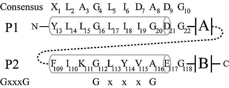
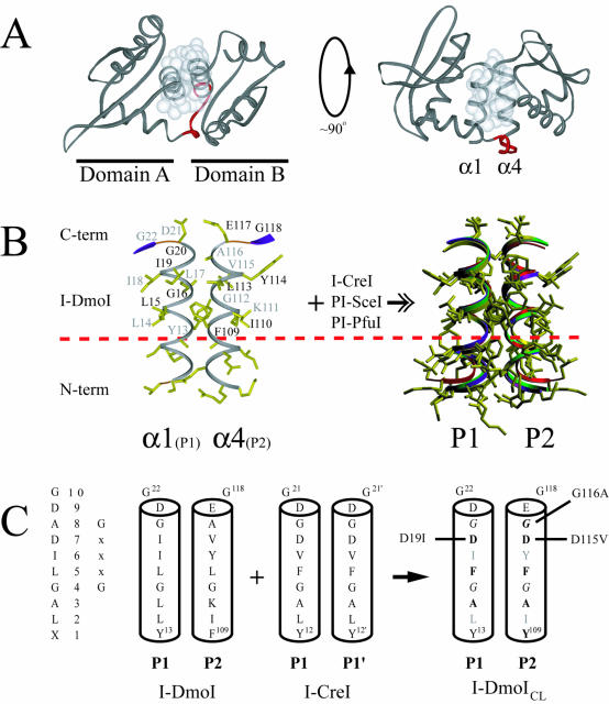
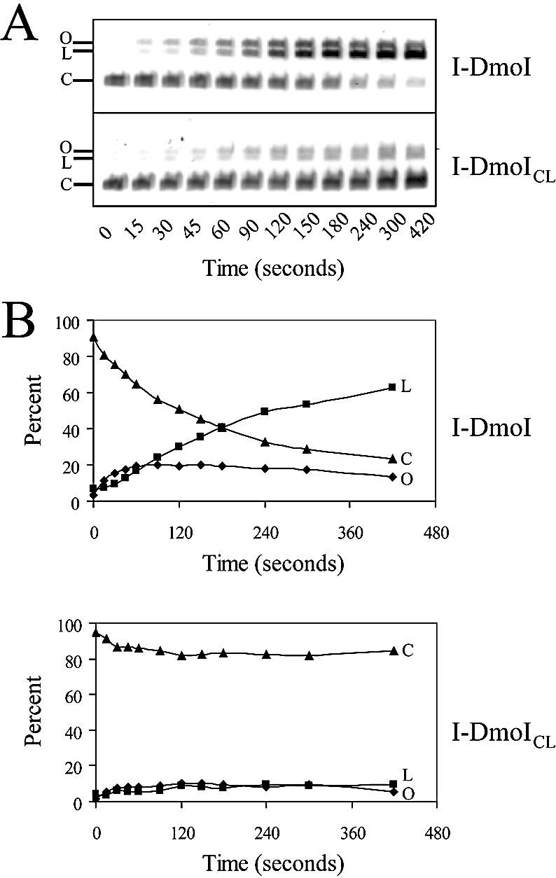
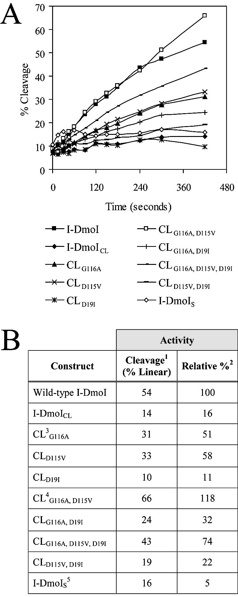
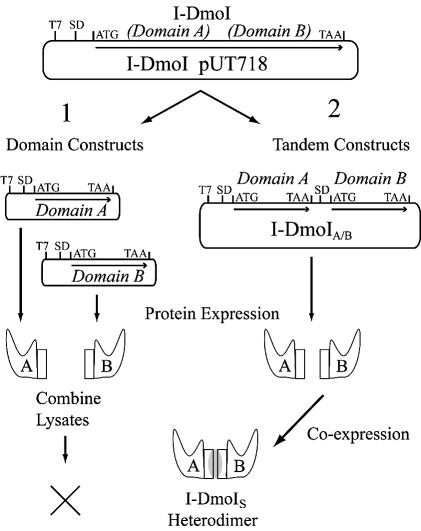
The general structural fold of the LAGLIDADG endonuclease family consists of two similar alpha/beta domains (alphabetabetaalphabetabetaalpha) that assemble either as homodimers or monomers with the domains related by pseudo-two-fold symmetry. At the center of this symmetry is the closely packed LAGLIDADG two-helix bundle that forms the main inter- or intra-molecular contact region between the domains of single- or double-motif proteins, respectively. In this work, we further examine the role of the LAGLIDADG residues involved in the helix-helix interaction. The interchangeability of the LAGLIDADG helix interaction was explored by grafting interfacial residues from the homodimeric I-CreI into the corresponding positions in the monomeric I-DmoI. The resulting LAGLIDADG exchange mutant is partially active, preferring to nick dsDNA rather than making the customary double-strand break. A series of partial revertants within the mutated LAGLIDADG region are shown to restore cleavage activity to varying degrees resulting in one I-DmoI mutant that is more active than wild-type I-DmoI. The phenotype of some of these mutants was reconciled on the basis of similarity to the GxxxG helix interaction found in transmembrane proteins. Additionally, a split variant of I-DmoI was created, demonstrating that the LAGLIDADG helices of I-DmoI are capable of forming and maintaining the protein-protein interface in trans to create an active heterodimer.


Figures

Figure 1
Figure 1
Schematic alignment of the LAGLIDADG residues of I-DmoI. Pertinent residues in P1 and P2 are identified below the consensus LAGLIDADG sequence. The alignment of the GxxxG motif is shown on the bottom. Numbers in the consensus sequences represent generic positions within the LAGLIDADG motif. Numbers in P1/P2 represent positions of residues in I-DmoI. Gray barrels indicate helical structure. A and B designate the two α/β domains that form the bulk of the protein. Dashed line represents the short (six to eight amino acids) linker between the subdomains A and B in I-DmoI.
Figure 2
Figure 2
Structure of I-DmoI and the LAGLIDADG motif. (A) Overall fold of I-DmoI. Left, top view down the two-helix bundle axis. Right, side view perpendicular to bundle axis. White spheres represent the van der Waals radii of the interfacial LAGLIDADG residues highlighting the majority of subdomain contacts between helices α1 and α4. Linker residues 98–105 are shown in red. (B) The α1/α4 helices (P1 and P2 regions) of I-DmoI with the XLAGLIDADG residues labeled. Black type, residues above the plane of the page; gray type, residues below the plane of the page. A dashed horizontal line demarcates the conserved sequence. At the right is the superposition of the LAGLIDADG helices of four known structures (I-DmoI, I-CreI, PI-SceI and PI-PfuI) demonstrating the extent of structural similarity in naturally occurring proteins. Although E-DreI, I-MsoI and I-AniI display a similar overlap, for clarity they were omitted from the superposition. (C) Creating I-DmoICL. The consensus sequence and generic numbering are shown on the left. The interfacial P1/P2 regions of I-DmoI were replaced by the P1/P1′ of I-CreI to generate I-DmoICL; bold type, residues changed in the LAGLIDADG of I-DmoI; gray type, residues facing the protein core and thus not changed to their I-CreI counterparts; italics, the GxxxG motif residues within the LAGLIDADG helices. Revertants are indicated to the left and right of the helices. See Figure 4 and the text for details.
Figure 3
Figure 3
Cleavage activity of I-DmoI and I-DmoICL. (A) Typical time-course activity assay. The 7 min assay was performed with pUD718 plasmid dsDNA and I-DmoI (top) or I-DmoICL (bottom); 20 µl samples were removed at the times indicated. Closed-circular plasmid DNA (C) was cleaved forming a linear product (L); nicked, open circles (O) appeared as intermediates of the reaction. Residual nuclease activity was ruled out by incubating each construct with target plasmid lacking only the 14 bp recognition sequence. No nicking or cleavage activity was observed (G. H. Silva, unpublished results). (B) Plots of data in (A). Percent reaction products are shown over time. Diamonds, squares and triangles correspond to O, L and C, respectively, in (A).
Figure 4
Figure 4
Relative cleavage activity of the I-DmoI constructs. (A) Time-course assays. Activity assays were performed in triplicate (average deviation <5%) as noted in Figure 3 for each construct listed below the plots. Percent cleavage reflects the ratio of linear to closed-circular dsDNA. (B) Relative cleavage activity. 1Cleavage activity on circular target, resulting in linear and nicked dsDNA; 2relative cleavage to wild-type I-DmoI based on the slope of the best fit line to each curve in (A); 3CL is I-DmoICL base construct with residue changes as indicated (see text for details); 4CLG116A, D115V is termed I-DmoIH in later experiments; 5I-DmoIS corresponds to split construct I-DmoI102/101
Figure 5
Figure 5
Creating a functional heterodimer. The wild-type I-DmoI plasmid was used as the template in efforts to create I-DmoIS via two pathways as shown. T7, promoter region; SD, Shine–Dalgarno sequence; ATG and TAA are the start and stop codons, respectively. Coding regions are indicated by arrows. (1) Domain constructs: individual A and B domain expression vectors were created. Protein expression was carried out separately for each domain and then lysates were combined and tested for heterodimer formation based on DNA cleavage activity. No active constructs were generated by this method. (2) Tandem constructs: individual A and B domain coding sequences were cloned into a single plasmid and co-expressed under the control of a single T7 promoter. Constructs were tested for heterodimer formation based on DNA cleavage activity as described in Table 4, Figure 6 and the text.
Figure 6
Figure 6
Cleavage-site mapping of I-DmoICL, I-DmoIH and I-DmoIS. The cleavage positions (arrows) on the top (left) and bottom (right) strand were mapped for I-DmoI (lane 1), I-DmoICL (lane 2), I-DmoIH (lane 3) and I-DmoIS (lane 4). Lane 5, no-protein control. A, C, G and T indicate the base in the sequencing ladder; left and right, the sequence readout for the area of interest as marked; bottom, the I-DmoI homing site with the cleavage position indicated.
Figure 7
Figure 7
The extent of heterodimer formation in I-DmoIS. (A) Schematic of the I-DmoI constructs used to probe heterodimer formation. I-DmoI constructs were created with the N-terminal FLAG (black square) and/or C-terminal c-myc (black circle) epitopes. Abbreviations for construct names appear in parenthesis to the left of each diagram. (B) Western blots quantifying I-DmoI and I-DmoIS. For all panels, sizes indicate expected position of wild-type I-DmoI (22 kDa) and split constructs (11 kDa; I-DmoIS and I-DmoISS). Panel 1 shows that I-DmoIS does not contain detectable full-length protein using anti-I-DmoI polyclonal antibody. Shaded triangles indicate decreasing protein concentration. Panel 2 shows that no cross-reactivity is evident for untagged or c-myc-tagged I-DmoI and I-DmoIS using the anti-FLAG antibody (the shadow band between lanes fW and Wc is an exposure artifact). Panel 3 shows the dramatically reduced amount of domain B in protein lysates (total protein amounts loaded are as in panel 2) when c-myc-tagged I-DmoI, I-DmoIS and I-DmoISS proteins were probed with anti-c-Myc antibody. Sc1 and Sc2 represent crude and cleared lysates of Sc protein, respectively. (C) Schematic of IP/western blot protocol. This method was used to determine relative dimerization. See text for details. (D) Western blot of dimerization. Results from the protocol outlined in (C). Panels 1a and 1b show different exposures of the same experiment; Panels 2a and 2b show the results of two separate experiments. Size markers are as in (B). IP indicates antibody used for immunoprecipitation: L, lysate-only control (no IP); F, anti-FLAG antibody; and C, anti-c-Myc antibody. Western indicates antibody used for western blot. By-products of the IP step: LCc, light chain of the anti-c-Myc antibody; LCf, light chain of the anti-FLAG antibody.

Similar articles

Cited by

References

    1. Dalgaard J.Z., Klar,A., Moser,M.J., Holley,W.R., Chatterjee,A. and Mian,I.S. (1997) Statistical modeling and analysis of the LAGLIDADG family of site-specific endonucleases and identification of an intein that encodes a site-specific endonuclease of the H-N-H family. Nucleic Acids Res., 25, 4626–4638. - PMC - PubMed
    1. Lucas P., Otis,C., Mercier,J.P., Turmel,M. and Lemieux,C. (2001) Rapid evolution of the DNA-binding site in LAGLIDADG homing endonucleases. Nucleic Acids Res., 29, 960–969. - PMC - PubMed
    1. Pietrokovski S. (1994) Conserved sequence features of inteins (protein introns) and their use in identifying new inteins and related proteins. Protein Sci., 3, 2340–2350. - PMC - PubMed
    1. Hensgens L.A.M., Bonen,L., de Haan,M., van der Horst,G. and Grivell,L.A. (1983) Two intron sequences in yeast mitochondrial COX1 gene: homology among URF-containing introns and strain-dependent variation in flanking exons. Cell, 32, 379–389. - PubMed
    1. Chevalier B.S. and Stoddard,B.L. (2001) Homing endonucleases: structural and functional insight into the catalysis of intron/intein mobility. Nucleic Acids Res., 29, 3757–3774. - PMC - PubMed

Publication types

Substances