

Human blood IgM "memory" B cells are circulating splenic marginal zone B cells harboring a prediversified immunoglobulin repertoire
- PMID: 15191950
- PMCID: PMC2590648
- DOI: 10.1182/blood-2004-01-0346
Human blood IgM "memory" B cells are circulating splenic marginal zone B cells harboring a prediversified immunoglobulin repertoire
Abstract
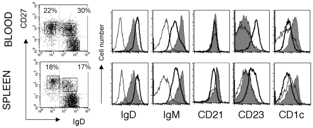
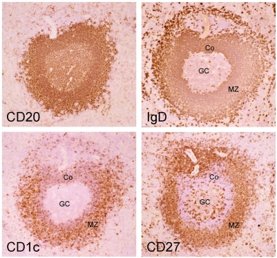
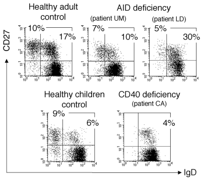
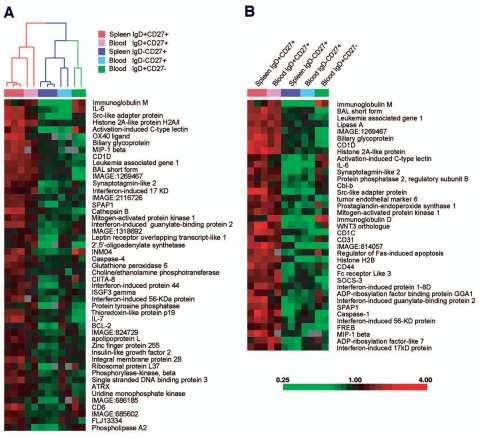
The human peripheral B-cell compartment displays a large population of immunoglobulin M-positive, immunoglobulin D-positive CD27(+) (IgM(+)IgD(+)CD27(+)) "memory" B cells carrying a mutated immunoglobulin receptor. By means of phenotypic analysis, complementarity-determining region 3 (CDR3) spectratyping during a T-independent response, and gene-expression profiling of the different blood and splenic B-cell subsets, we show here that blood IgM(+)IgD(+)CD27(+) cells correspond to circulating splenic marginal zone B cells. Furthermore, analysis of this peripheral subset in healthy children younger than 2 years shows that these B cells develop and mutate their immunoglobulin receptor during ontogeny, prior to their differentiation into T-independent antigen-responsive cells. It is therefore proposed that these IgM(+)IgD(+)CD27(+) B cells provide the splenic marginal zone with a diversified and protective preimmune repertoire in charge of the responses against encapsulated bacteria.
Figures
Similar articles
-
Somatic diversification in the absence of antigen-driven responses is the hallmark of the IgM+ IgD+ CD27+ B cell repertoire in infants.J Exp Med. 2008 Jun 9;205(6):1331-42. doi: 10.1084/jem.20071555. Epub 2008 Jun 2. J Exp Med. 2008. PMID: 18519648 Free PMC article.
-
Differential Expression of IgM and IgD Discriminates Two Subpopulations of Human Circulating IgM+IgD+CD27+ B Cells That Differ Phenotypically, Functionally, and Genetically.Front Immunol. 2020 May 6;11:736. doi: 10.3389/fimmu.2020.00736. eCollection 2020. Front Immunol. 2020. PMID: 32435242 Free PMC article.
-
Human immunoglobulin (Ig)M+IgD+ peripheral blood B cells expressing the CD27 cell surface antigen carry somatically mutated variable region genes: CD27 as a general marker for somatically mutated (memory) B cells.J Exp Med. 1998 Nov 2;188(9):1679-89. doi: 10.1084/jem.188.9.1679. J Exp Med. 1998. PMID: 9802980 Free PMC article.
-
[IgM+IgD+CD27+ B cells in human: an essential role in the protection against encapsulated bacteria].Med Sci (Paris). 2015 Jun-Jul;31(6-7):647-53. doi: 10.1051/medsci/20153106018. Epub 2015 Jul 7. Med Sci (Paris). 2015. PMID: 26152169 Review. French.
-
Memory B cells and CD27.Histol Histopathol. 2000 Apr;15(2):573-6. doi: 10.14670/HH-15.573. Histol Histopathol. 2000. PMID: 10809378 Review.
Cited by
-
Toll-like receptor signaling in primary immune deficiencies.Ann N Y Acad Sci. 2015 Nov;1356(1):1-21. doi: 10.1111/nyas.12763. Epub 2015 Apr 30. Ann N Y Acad Sci. 2015. PMID: 25930993 Free PMC article. Review.
-
IgM memory B cells: a mouse/human paradox.Cell Mol Life Sci. 2012 May;69(10):1625-34. doi: 10.1007/s00018-012-0971-z. Epub 2012 Apr 6. Cell Mol Life Sci. 2012. PMID: 22481437 Free PMC article. Review.
-
FCRL3 promotes TLR9-induced B-cell activation and suppresses plasma cell differentiation.Eur J Immunol. 2013 Nov;43(11):2980-92. doi: 10.1002/eji.201243068. Epub 2013 Aug 12. Eur J Immunol. 2013. PMID: 23857366 Free PMC article.
-
Gut microbiota and atherosclerosis: role of B cell for atherosclerosis focusing on the gut-immune-B2 cell axis.J Mol Med (Berl). 2020 Sep;98(9):1235-1244. doi: 10.1007/s00109-020-01936-5. Epub 2020 Jul 31. J Mol Med (Berl). 2020. PMID: 32737524 Free PMC article. Review.
-
PID comes full circle: applications of V(D)J recombination excision circles in research, diagnostics and newborn screening of primary immunodeficiency disorders.Front Immunol. 2011 May 4;2:12. doi: 10.3389/fimmu.2011.00012. eCollection 2011. Front Immunol. 2011. PMID: 22566803 Free PMC article.
References
-
- Klein U, Kuppers R, Rajewsky K. Evidence for a large compartment of IgM-expressing memory B cells in humans. Blood. 1997;89:1288–1298. - PubMed
-
- van Es JH, Meyling FH, Logtenberg T. High frequency of somatically mutated IgM molecules in the human adult blood B cell repertoire. Eur J Immunol. 1992;22:2761–2764. - PubMed
-
- Kumararatne DS, Bazin H, MacLennan IC. Marginal zones: the major B cell compartment of rat spleens. Eur J Immunol. 1981;11:858–864. - PubMed
Publication types
MeSH terms
Substances
LinkOut - more resources
Full Text Sources
Other Literature Sources
Research Materials
