. 2004 Jun 22;101(25):9411-6.
doi: 10.1073/pnas.0403160101. Epub 2004 Jun 10.

Herpes simplex virus protein kinase US3 activates and functionally overlaps protein kinase A to block apoptosis

Affiliations

Herpes simplex virus protein kinase US3 activates and functionally overlaps protein kinase A to block apoptosis

Luca Benetti et al. Proc Natl Acad Sci U S A. .

Abstract

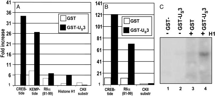
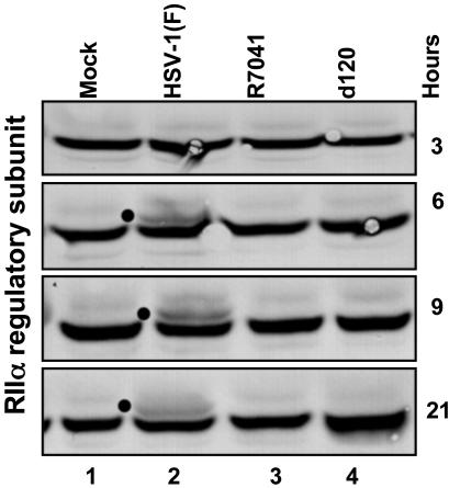
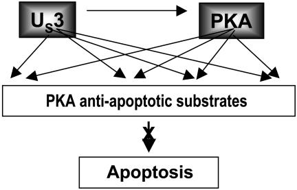
Herpes simplex virus 1 encodes at least four genes whose functions include blocking apoptosis induced by exogenous agents (e.g., sorbitol, Fas ligand, and BAD protein) or replication-incompetent mutants (e.g., the d120 mutant lacking both copies of the alpha 4 gene). U(S)3, one of these four genes, encodes a serine-threonine kinase that has been demonstrated to block apoptosis induced by proapoptotic cellular proteins or by the d120 mutant. The amino acid context of serine-threonine phosphorylated by U(S)3 is similar to that of the cAMP-dependent protein kinase PKA. We report that (i) the pattern of proteins phosphorylated by U(S)3 in transduced cells or in cells infected with WT virus overlaps that of phosphoproteins targeted by PKA, (ii) activation of PKA blocks apoptosis induced by d120 mutant or by BAD protein independently of U(S)3, (iii) U(S)3 protein kinase phosphorylates peptides containing the serine or threonine targeted by PKA including that present in the regulatory type II alpha subunit of PKA, and (iv) in WT virus-infected cells the regulatory type II alpha subunit is phosphorylated in a U(S)3-dependent manner. We conclude that a major determinant of the antiapoptotic activity of the U(S)3 protein kinase is the phosphorylation of PKA substrates by either or both enzymes.


Figures

Fig. 1.
Fig. 1.
Profiles of electrophoretically separated proteins reacted with antibody specific for phosphorylated serines or threonines in the context of sequences recognized by PKA. SK-N-SH cells were harvested 18 h after mock infection or infection with HSV-1(F), the recombinant virus R7041(ΔUS3) or d120(Δα4), or baculovirus infection. The electrophoretically separated proteins in a denaturing polyacrylamide gel were transferred to a nitrocellulose sheet and reacted with the antibody as described in Materials and Methods.
Fig. 2.
Fig. 2.
The effect of forskolin on the DEVDase activity of cells infected with the HSV-1 d120 mutant or transduced with a baculovirus encoding gstBAD3S/A chimeric protein. (A) HEp-2 cells were either mock-infected or infected with d120 mutant (10 pfu per cell) in the presence of increasing concentrations of forskolin. Cells were harvested 18 h after infection, and the cell lysates were assayed for DEVDase activity. (B) SK-N-SH cells were either mock-infected or infected with d120 mutant (10 pfu per cell) in the presence of 100 μM forskolin. Cells were harvested 18 h after the infection and assayed for DEVDase activity. (C) Rabbit skin cells were either mock-infected or transduced with a baculovirus expressing gstBAD3S/A (10 pfu per cell), in the presence or absence of 100 μM forskolin. Cells were harvested 18 h after infection and assayed for DEVDase activity. In lanes 4 and 5, cells were exposed to either Bac-WT or Bac-US3 for 6 h, superinfected with Bac-gstBAD3S/A for 18 h, lysed, and assayed for DEVDase activity.
Fig. 3.
Fig. 3.
The effect of forskolin on the cleavage of PARP and the synthesis of viral proteins in cells infected with the d120 mutant. HEp-2 cells mock-infected or infected with the d120 mutant (10 pfu per cell) in the presence or absence of 100 μM forskolin were harvested 18 h after infection, solubilized, subjected to electrophoresis in a denaturing polyacrylamide gel, transferred to a nitrocellulose sheet, and reacted with anti-PARP antibody (A) and antibody to infected-cell protein (ICP) no. 0 (B), ICP22 (C), or ICP27 (D).
Fig. 4.
Fig. 4.
DEVDase activity of cells infected with the HSV-1 d120 mutant in the presence or absence of forskolin or PKI. (A) Replicate cultures of HEp-2 cells were incubated for 90 min in medium containing 100 μM PKI. The cells were then either mock-infected, exposed to the d120 mutant (10 pfu per cell), or infected with the d120 mutant and treated with 100 μM forskolin. (B) Replicate cultures of HEp-2 cells were incubated for 90 min in the presence of increasing concentrations of PKI. The cells were then either mock-infected, infected with d120, or infected with d120 and treated with forskolin. In both sets of experiments, the cells were harvested 18 h after the infection, and the cell lysates were assayed for DEVDase activity.
Fig. 5.
Fig. 5.
Phosphorylation of known PKA substrates by GST-US3 kinase. Two micrograms (A)or4 μg(B) of the indicated peptides were reacted with 1 μg of purified GST or GST-Us3 attached to glutathione Sepharose beads, as described in the text. Phosphorylated substrates were adsorbed onto cellulose phosphate paper filters, and radioactivity was measured with a scintillation counter. The data show the increase in cpm over a mock reaction carried out with no substrate. (C) Four micrograms of histone H1 were reacted with 1 μg of purified GST or GST-US3 attached to glutathione Sepharose beads, as described in the text. Samples were subjected to polyacrylamide gel electrophoresis, nitrocellulose membrane transfer, and autoradiography.
Fig. 6.
Fig. 6.
Posttranslational modification of PKA RIIα in infected cells requires the presence of US3 protein kinase. HEp-2 cells were either mock-infected or infected with HSV-1(F) or the R7041 or d120 mutant viruses (10 pfu per cell). The cells were harvested at 3, 6, 9, or 21 h after infection. Cell lysates were separated on an 8% denaturing polyacrylamide gel, transferred to a nitrocellulose sheet, and reacted with antibody against the RIIα subunit of PKA. The dots identify the appearance of posttranslationally modified forms in infected cells.
Fig. 7.
Fig. 7.
A model for the antiapoptotic interplay between US3 and PKA. Earlier studies have shown that the US3 protein kinase blocks apoptosis induced by d120, a replication-incompetent virus, and by ectopically expressed BAD protein. This report shows that activated PKA blocks apoptosis induced by either d120 mutant or BAD protein, and that US3 can phosphorylate PKA substrates including the autophosphorylation site of the RIIα subunit of PKA. The model predicts that the US3 protein kinase activates PKA, and that either PKA or both PKA and US3 enzymes block apoptosis.

Similar articles

Cited by

References

    1. Galvan, V. & Roizman, B. (1998) Proc. Natl. Acad. Sci. USA 95, 3931-3936. - PMC - PubMed
    1. Jerome, K. R., Chen, Z., Lang, R., Torres, M. R., Hofmeister, J., Smith, S., Fox, R., Froelich, C. J. & Corey, L. (2001) J. Immunol. 167, 3928-3935. - PubMed
    1. Jerome, K. R., Fox, R., Chen, Z., Sears, A. E., Lee, H.-Y. & Corey, L. (1999) J. Virol. 73, 8950-8957. - PMC - PubMed
    1. Sieg, S., Yildirim, Z., Smith, D., Kayagaki, N., Yagita, H., Huang, Y. & Kaplan, D. (1996) J. Virol. 70, 8747-8751. - PMC - PubMed
    1. Galvan, V., Brandimarti, R. & Roizman, B. (1999) J. Virol. 73, 3219-3226. - PMC - PubMed

Publication types

MeSH terms

Substances