. 2004 Jul 7;23(13):2586-96.
doi: 10.1038/sj.emboj.7600251. Epub 2004 Jun 10.

PS1 activates PI3K thus inhibiting GSK-3 activity and tau overphosphorylation: effects of FAD mutations

Affiliations

PS1 activates PI3K thus inhibiting GSK-3 activity and tau overphosphorylation: effects of FAD mutations

Lia Baki et al. EMBO J. .

Abstract

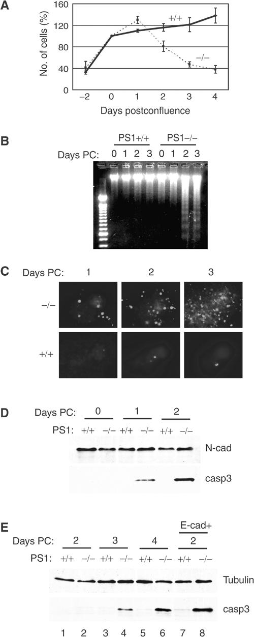
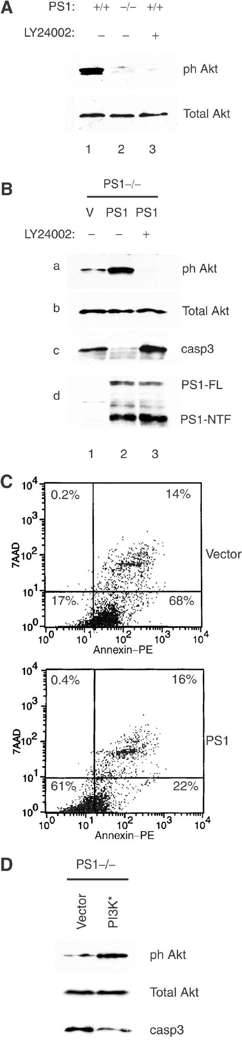
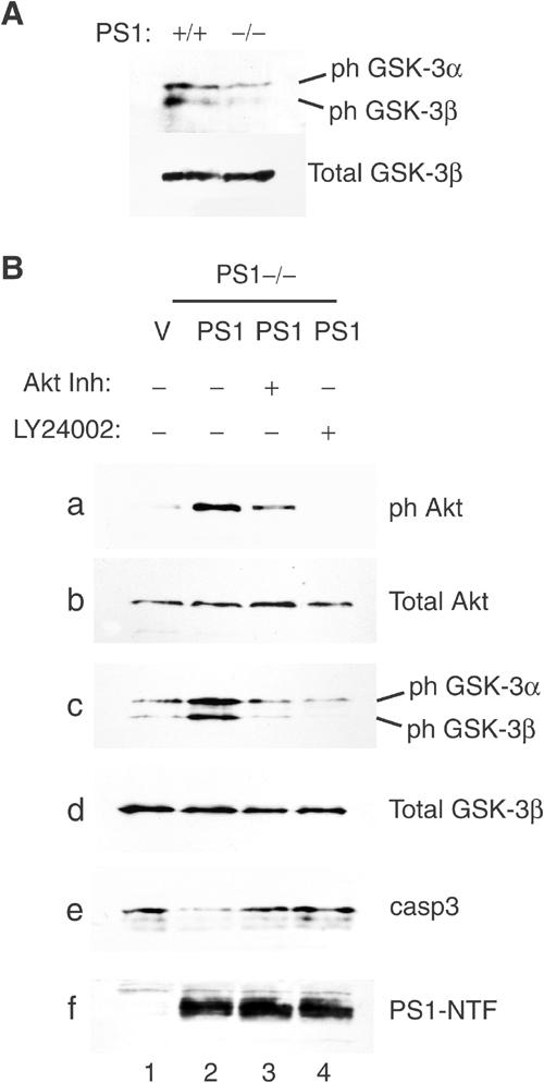
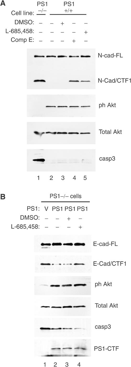
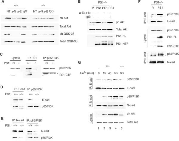
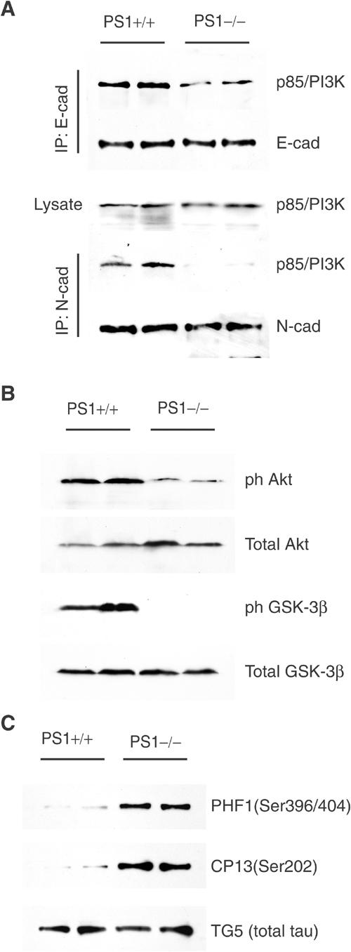
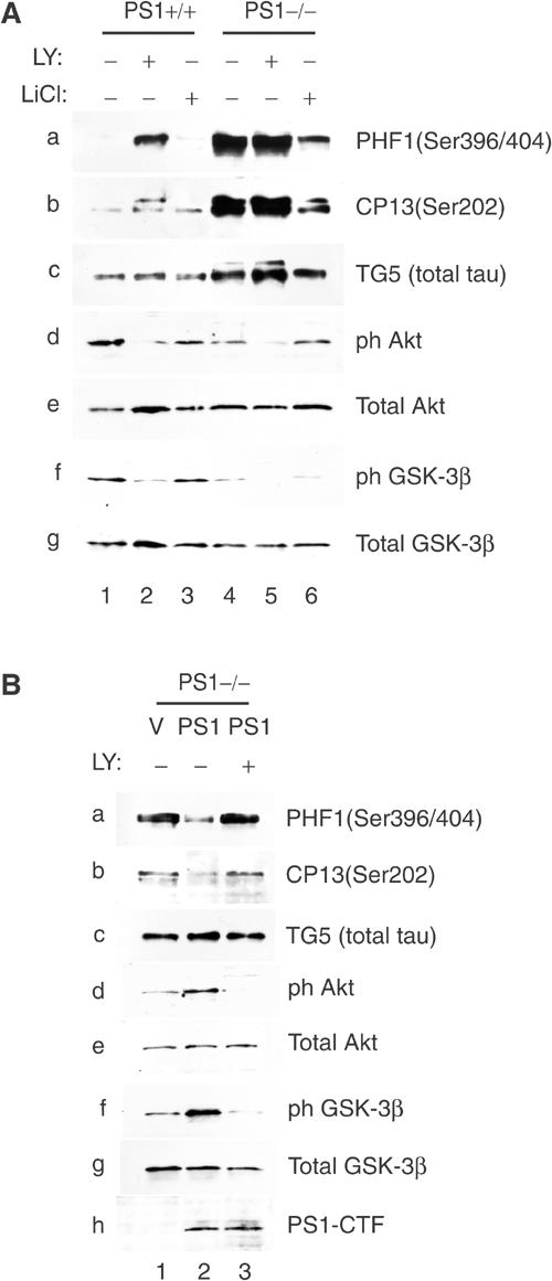
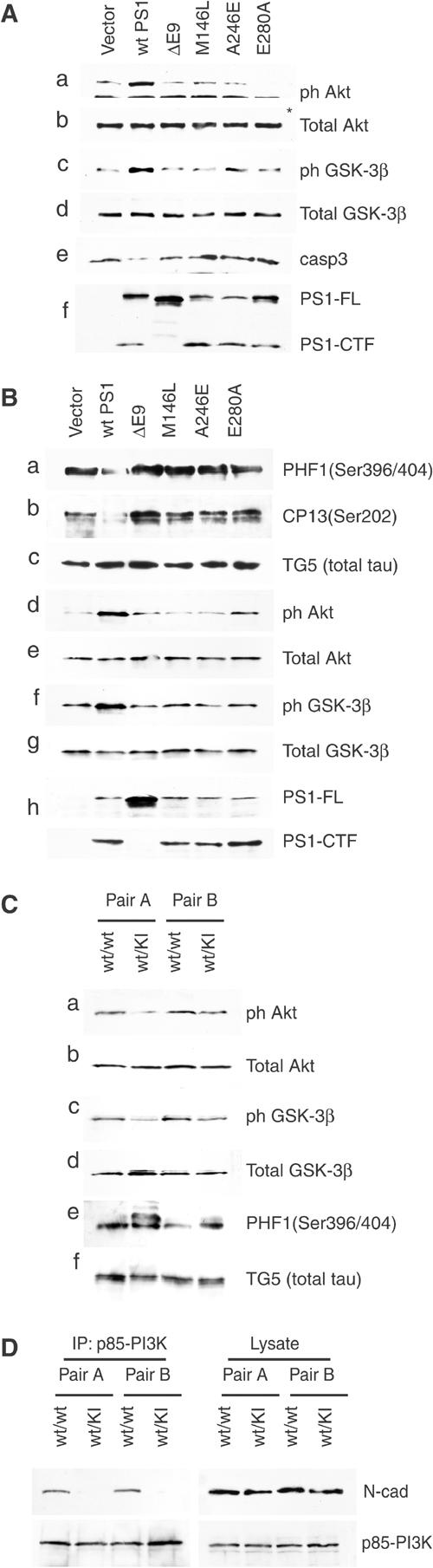
Phosphatidylinositol 3-kinase (PI3K) promotes cell survival and communication by activating its downstream effector Akt kinase. Here we show that PS1, a protein involved in familial Alzheimer's disease (FAD), promotes cell survival by activating the PI3K/Akt cell survival signaling. This function of PS1 is unaffected by gamma-secretase inhibitors. Pharmacological and genetic evidence indicates that PS1 acts upstream of Akt, at or before PI3K kinase. PS1 forms complexes with the p85 subunit of PI3K and promotes cadherin/PI3K association. Furthermore, conditions that inhibit this association prevent the PS1-induced PI3K/Akt activation, indicating that PS1 stimulates PI3K/Akt signaling by promoting cadherin/PI3K association. By activating PI3K/Akt signaling, PS1 promotes phosphorylation/inactivation of glycogen synthase kinase-3 (GSK-3), suppresses GSK-3-dependent phosphorylation of tau at residues overphosphorylated in AD and prevents apoptosis of confluent cells. PS1 FAD mutations inhibit the PS1-dependent PI3K/Akt activation, thus promoting GSK-3 activity and tau overphosphorylation at AD-related residues. Our data raise the possibility that PS1 may prevent development of AD pathology by activating the PI3K/Akt signaling pathway. In contrast, FAD mutations may promote AD pathology by inhibiting this pathway.


Figures

Figure 1
Figure 1
Absence of PS1 triggers density-dependent apoptosis. (A) Survival analysis of E-cadherin-transfected PS1+/+ or PS1−/− cells (continuous and dotted lines, respectively) cultured up to 4 days postconfluence. Mean values, ±s.e.m., from two independent experiments (each in triplicate) are presented. (B) Kinetics of the appearance of apoptotic (fragmented) DNA in nuclei of E-cadherin-transfected PS1+/+ and PS1−/− cells, cultured for up to 3 days postconfluence (Days PC). (C) Kinetics of the appearance of TUNEL-positive (apoptotic) nuclei in E-cadherin-transfected PS1+/+ and PS1−/− cells, cultured for up to 3 days postconfluence. (D) Lysates from 0-, 1- and 2-day postconfluent E-cadherin-transfected PS1+/+ and PS1−/− cells were analyzed with anti-N-cadherin (loading control) and anti-activated (cleaved) caspase-3 antibodies. N-cad: N-cadherin; casp3: activated caspase-3. (E) Lysates from 2- to 4-day postconfluent nontransfected PS1+/+ and PS1−/− cells (lanes 1–6) and from 2-day postconfluent E-cadherin-transfected PS1+/+ and PS1−/− cells (lanes 7 and 8) were analyzed with anti-b-tubulin (control) and anti-activated (cleaved) caspase-3 antibodies.
Figure 2
Figure 2
PS1 inhibits apoptosis by activating PI3K/Akt signaling. (A) Lysates from confluent E-cadherin-transfected PS1+/+ and PS1−/− fibroblasts, cultured in the absence or presence of the PI3K inhibitor LY24002, were analyzed for phosphorylated Akt at Ser473 (ph Akt) or total Akt. (B) PS1−/− cells infected with HSV vector (V) or HSV PS1 (PS1) were cultured in the presence or absence of PI3K inhibitor (LY24002). Lysates were analyzed as shown. ph Akt: phosphorylated Akt at Ser473; casp3: activated caspase-3; PS1-FL: full-length PS1; PS1-NTF: NTF fragment of PS1. (C) PS1−/− cells, transiently transfected either with EGFP vector (V) or with EGFP-PS1 (PS1), were subjected to annexin/7AAD staining and analyzed by flow cytometry. Only the EGFP-positive (transfected) cells are shown. Percentages refer to total EGFP-positive cells. Nonapoptotic cells are shown in the lower left square. A total of 10 000 EGFP-positive cells were analyzed. (D) PS1−/− cells were transfected with mutant (active) PI3K (PI3K*) or empty vector and lysates were analyzed as shown.
Figure 3
Figure 3
PS1 regulates GSK-3 activity via the PI3K/Akt pathway. (A) Lysates from confluent PS1+/+ or PS1−/− fibroblasts were analyzed for phosphorylation of GSK-3α (ph GSK-3α) and GSK-3β (ph GSK-3β) at Ser21 and Ser9, respectively. (B) PS1−/− cells infected with HSV vector (V) or HSV PS1 (PS1) were cultured in the presence or absence of either Akt inhibitor (Akt Inh) or PI3K inhibitor (LY24002). Lysates were analyzed as shown. PS1-NTF: NTF fragment of PS1.
Figure 4
Figure 4
PS1-mediated activation of the PI3K–Akt pathway is independent of γ-secretase activity. (A) Confluent PS1−/− (lane 1) or PS1+/+ cells (lanes 2–5) were cultured overnight in the presence or absence of γ-secretase inhibitor XVIII (Compound E, lane 4), γ-secretase inhibitor L-685,458 (lane 5) or vehicle (DMSO, lane 3). Lysates were analyzed as shown. N-cad-FL: full-length N-cadherin; N-cad-CTF1: CTF1 fragment of N-cadherin (Marambaud et al, 2003). (B) PS1−/− cells were infected with HSV vector (V) or HSV PS1 (PS1) and cultured in the presence or absence of γ-secretase inhibitor. Lysates were analyzed as shown. E-cad-FL: full-length E-cadherin; E-cad-CTF1: CTF1 fragment of E-cadherin (Marambaud et al, 2002); PS1-CTF: CTF fragment of PS1.
Figure 5
Figure 5
PS1 activates the PI3K/Akt pathway by promoting association of the p85 subunit of PI3K with E- and N-cadherin. (A) Postconfluent PS1+/+ or PS1−/− cells were cultured in the presence of anti-N-cadherin and anti-E-cadherin antibodies (a-N and a-E, respectively) or isotypic IgG (IgG). Lysates were analyzed as shown. NT: no treatment. (B) PS1−/− cells were infected with HSV vector (V) or HSV PS1 (PS1) and cultured in the presence of both anti-E- and anti-N-cadherin antibodies (anti-E+anti-N) or isotypic IgG (IgG). Lysates were analyzed as shown. (C) Lysates from postconfluent PS1+/+ or PS1−/− cells were immunoprecipitated (IPed) with anti-PS1 (IP: PS1) or anti-p85/PI3K (IP: p85/PI3K) antibodies and obtained immunoprecipitates (IPs) were analyzed with anti-p85/PI3K or anti-PS1-CTF antibodies as shown. Asterisk denotes IgG. (D) Lysates from postconfluent PS1+/+ or PS1−/− cells were IPed with anti-E-cadherin or anti-p85/PI3K antibodies and obtained IPs were analyzed for E-cadherin or p85/PI3K. (E) Lysates as in (D) were IPed with anti-N-cadherin or anti-p85/PI3K antibodies and obtained IPs were analyzed as shown. (F) PS1−/− cells were infected with HSV vector (V) or HSV PS1 (PS1). Lysates were IPed with anti-E- or anti-N-cadherin antibodies and analyzed as shown. Asterisk denotes IgG. (G) Confluent PS1+/+ cells were preincubated for 45 min in the presence of 4 mM EGTA to break cadherin/PI3K complexes (Pece et al, 1999; Tran et al, 2002) and then cultures were switched to Ca2+-containing medium at time zero and followed for the times shown (lanes 1–3). Lysates were IPed with anti-E- or anti-N-cadherin antibodies and IPs were analyzed with anti-p85/PI3K, or anti-E- and anti-N-cadherin antibodies as shown. Steady-state (SS) levels of cadherin/PI3K complexes in the absence of EGTA were monitored in PS1+/+ and PS1−/− cultures (lanes 4 and 5, respectively). All cultures were in serum-free media.
Figure 6
Figure 6
PS1 knockout embryos show reduced cadherin/PI3K complexes, decreased phosphorylation of Akt and GSK-3 and increased GSK-3-dependent phosphorylation of tau. (A) Total embryo homogenates prepared from PS1+/+ or PS1−/− mouse embryo littermates were immunoprecipitated with anti-E-cadherin (IP: E-cad) or anti-N-cadherin (IP: N-cad) antibodies and analyzed as shown. (B) Lysates were prepared from PS1+/+ or PS1−/− embryonic brains and analyzed for phosphorylated Akt and GSK-3β as shown. (C) Lysates were prepared from PS1+/− and PS1−/− mouse embryonic brains. The heat-stable fraction of lysates was analyzed with phosphorylation-dependent (PHF1, CP13) and phosphorylation-independent (TG5) anti-tau antibodies. Duplicate samples each from a littermate embryo are shown.
Figure 7
Figure 7
PS1 controls tau phosphorylation via the PI3K/Akt/GSK-3β pathway. (A) PS1+/+ or PS1−/− fibroblasts transiently expressing the longest human tau isoform were cultured in the presence or absence of PI3K inhibitor LY24002 (LY) or GSK-3 inhibitor LiCl. Lysates were analyzed for phosphorylation of tau (panels a–c), phosphorylation of Akt (panels d and e) and phosphorylation of GSK-3β (panels f and g). Transfected tau is detected by anti-tau antibody TG5 (panel c, total tau). (B) Human tau-expressing PS1−/− fibroblasts (see A) were infected with HSV vector (V) or HSV PS1 (PS1) and cultured in the presence or absence of PI3K inhibitor LY24002. Lysates were analyzed on Western blots as shown.
Figure 8
Figure 8
PS1 FAD mutants are defective in their ability to activate the PI3K–Akt pathway and to suppress phosphorylation of tau. (A) E-cadherin-expressing PS1−/− fibroblasts were infected with HSV vector (vector), with HSV recombinant viruses encoding WT PS1 (wt PS1) or with one of the PS1 FAD mutations shown on the top of each lane. Lysates prepared at 40 h postinfection were analyzed for PS1 expression (panel f), Akt phosphorylation (Ser473, panels a and b), GSK-3β phosphorylation (Ser9, panels c and d) or activated caspase-3 (panel e). Asterisk denotes nonspecific signal. (B) E-cadherin-expressing PS1−/− cells transiently expressing the longest human tau isoform were infected with HSV vector (vector), with HSV recombinant viruses encoding WT PS1 (wt PS1) or with one of the PS1 FAD mutants shown on the top of each lane. Lysates were analyzed for PS1 expression (panel h), tau phosphorylation (panels a–c) and phosphorylation of Akt and GSK-3β (panels d–g). (C) Brain lysates were prepared from two independent pairs of mice each pair consisting of a heterozygous PS1 I213T knock-in mouse (Nakano et al, 1999) and its WT littermate. Lysates were analyzed for phosphorylation of Akt (panels a and b), GSK-3β (panels c and d) and tau (panels e and f). Increased levels of multiple isoforms of overphosphorylated tau are detected in the knock-in mice. (D) Brain homogenates from two independent pairs of heterozygous PS1 I213T knock-in mice and their WT littermates (see C) were immunoprecipitated with anti-p85/PI3K antibodies and analyzed as shown.

Similar articles

Cited by

References

    1. Baki L, Marambaud P, Efthimiopoulos S, Georgakopoulos A, Wen P, Cui W, Shioi J, Koo E, Ozawa M, Friedrich VL Jr, Robakis NK (2001) Presenilin-1 binds cytoplasmic epithelial cadherin, inhibits cadherin/p120 association, and regulates stability and function of the cadherin/catenin adhesion complex. Proc Natl Acad Sci USA 98: 2381–2386 - PMC - PubMed
    1. Brunet A, Datta SR, Greenberg ME (2001) Transcription-dependent and -independent control of neuronal survival by the PI3K–Akt signaling pathway. Curr Opin Neurobiol 11: 297–305 - PubMed
    1. Bursztajn S, DeSouza R, McPhie DL, Berman SA, Shioi J, Robakis NK, Neve RL (1998) Overexpression in neurons of human presenilin-1 or a presenilin-1 familial Alzheimer disease mutant does not enhance apoptosis. J Neurosci 18: 9790–9799 - PMC - PubMed
    1. Chan TO, Rittenhouse SE, Tsichlis PN (1999) AKT/PKB and other D3 phosphoinositide-regulated kinases: kinase activation by phosphoinositide-dependent phosphorylation. Annu Rev Biochem 68: 965–1014 - PubMed
    1. Chan TO, Tsichlis PN (2001) PDK2: a complex tail in one Akt. Sci STKE 2001: PE1. - PubMed

Publication types

MeSH terms