Comparative Study
. 2004 Jul 7;23(13):2498-509.
doi: 10.1038/sj.emboj.7600263. Epub 2004 Jun 10.

Interactions between UvrA and UvrB: the role of UvrB's domain 2 in nucleotide excision repair

Affiliations
Comparative Study

Interactions between UvrA and UvrB: the role of UvrB's domain 2 in nucleotide excision repair

James J Truglio et al. EMBO J. .

Abstract

Nucleotide excision repair (NER) is a highly conserved DNA repair mechanism present in all kingdoms of life. UvrB is a central component of the bacterial NER system, participating in damage recognition, strand excision and repair synthesis. None of the three presently available crystal structures of UvrB has defined the structure of domain 2, which is critical for the interaction with UvrA. We have solved the crystal structure of the UvrB Y96A variant, which reveals a new fold for domain 2 and identifies highly conserved residues located on its surface. These residues are restricted to the face of UvrB important for DNA binding and may be critical for the interaction of UvrB with UvrA. We have mutated these residues to study their role in the incision reaction, formation of the pre-incision complex, destabilization of short duplex regions in DNA, binding to UvrA and ATP hydrolysis. Based on the structural and biochemical data, we conclude that domain 2 is required for a productive UvrA-UvrB interaction, which is a pre-requisite for all subsequent steps in nucleotide excision repair.


Figures

Figure 1
Figure 1
Three-dimensional structure of the UvrA-interacting domain (domain 2) of UvrB. The ribbon diagram shows the secondary structure elements and mutated residues on the proposed UvrA interacting face. The core β sheet (β2–β7) is shown in green, a second sheet in blue (β1, β8) and the single α helix in pink. Blue spheres as well as residue labels mark the beginning and end of domain 2.
Figure 2
Figure 2
Sequence conservation of domain 2 in UvrB. (A) Surface representation of domain 2 of UvrB (gray) with conserved residues labeled and color-coded (red: strictly conserved, dark blue: very highly conserved, cyan: highly conserved, green: moderate to highly conserved). Conservation is based on 56 domain 2 sequences aligned using ClustalX and analyzed by the ConSurf server. The upper panel shows the front (DNA-binding) side of UvrB and the lower panel is a 180° rotation showing the back. The remainder of UvrB is drawn as a Cα trace and color-coded according to domain architecture with domain 1a in yellow, 1b in green, 3 in red and the β hairpin in cyan. (B) Sequence alignment of UvrB domain 2 (first block) and the homologous domain in Mfd (second block). UvrB sequences from Bacillus burgdorferi (gi:8134783), Helicobacter pylori (gi:15645728), T. thermophilus (gi:2499102), Mycoplasma genitalium (gi:12044925), Staphylococcus aureus subsp. aureus MW2 (gi:21282449), E. coli (gi 137190), Salmonella typhimurium species LT2 (gi 16764161) and Mycobacterium tuberculosis (gi 3122992), and Mfd sequences from E. coli strain 0157:H7 (gi:15830746), Yersinia pestis (gi:16121893), B. burgdorferi (gi:3914012), Corynebacterium glutamicum species ATCC 13032 (gi:41325189), M. tuberculosis species H37Rv (gi:15608160), S. aureus subsp. aureus N315 (gi:15926180) and Chlamydia trachomatis (gi:15605481) are included. The secondary structure is indicated above the sequence with arrows for β-strands and a cylinder for the α-helix. Color coding of secondary structure was chosen to match that of Figure 1. Residues are highlighted according to the conservation shown in (A).
Figure 3
Figure 3
Comparison of the Y96A UvrB structure to WT UvrB. (A) Stereo view of the interface between domain 2 and the remainder of the UvrB molecule. Selected side chains are shown and labeled. Color coding is according to domain architecture as in Figure 2A and domain 2 in blue. Hydrogen bonds and salt bridges are indicated by red dotted lines. (B) Comparison of the overall structure of WT UvrB (cyan) and the two NCS-related copies of UvrB Y96A (yellow and red) as a stereo view. Orientation is chosen as in Figure 2. For the superposition, domain 1a of each of the structures was used and the resulting transformations were applied to the entire molecule. (C) Superposition of UvrB Y96A (color coded as in Figure 2) and WT UvrB (gray). Side chains for Tyr 92, Asp 117 and Arg 190 are shown for both the WT and the UvrB Y96A structure. The side chain of Y96 is omitted from the native model since the electron density for this residue is insufficient. A sphere indicates the position of the Cα atom of Y96 (A96 for the mutant).
Figure 4
Figure 4
Incision activity of domain 2 mutants. (A, B, C): The 5′-end-labeled substrate was incubated with 20 nM UvrA, 50 nM UvrC and 100 nM of the indicated UvrB protein for 5 min (A) or 30 min (B, C), at 55°C in reaction buffer. The reactions were terminated with stop buffer, and the incision products were analyzed on a 10% denaturing polyacrylamide gel. (D, E): Comparison of the incision activity at 5 min (black bars) and 30 min (white bars) using the indicated UvrB proteins. Data are reported as the mean±the standard deviation of the mean of two to four incision assays per time point and substrate. Panels A, B and D: 50 mer dsDNA substrate containing a centrally located fluorescein (FldT). Panels C and E: 50 mer dsDNA substrate containing a centrally located single-nucleotide gap.
Figure 5
Figure 5
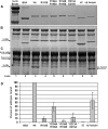
UvrA pull-down assay. The UvrA-chitin beads were incubated with either WT UvrB or the mutants. After washing the beads extensively, the bound proteins were analyzed on a 10% denaturing polyacrylamide gel. (A) Sample of all proteins used in the study. (B) One-twentieth of the reactions, the ‘inputs'. (C) Proteins that remained bound to the resin after extensive washing. (D) Quantitation of panel C reporting the percent of WT UvrB bound (data reported as the mean±the standard error of the mean n=2). The asterisk (*) in panel C, lane 1 indicates a nonspecific band observed in all reaction lanes, which migrates just above the band for UvrB. For quantitation, the area of this band was subtracted from all lanes except Δ2 and Δβ hairpin, whose proteins migrate faster.
Figure 6
Figure 6
Protein–DNA complex formations by UvrA and WT UvrB or UvrB mutants. UvrA (20 nM) was incubated with the various UvrB proteins (120 nM) as indicated at 55°C for 5 min (A) or 20 min (B) in the presence of 2 nM F26, 50 duplex DNA with the modified strand 5′ terminally labeled. The reaction mixtures were analyzed on 4% polyacrylamide native gels in the presence of 1 mM ATP and 10 mM MgCl2. (C) Quantitation of EMSAs in panels A and B, reporting the percent of DNA bound to UvrA, WT UvrB or UvrB mutants at 5 and 20 min (data are reported as the mean±the standard deviation (n=3) for each time point). White bars (solid or striped) indicate the percentage of DNA bound as the AB:DNA/A2:DNA or B:DNA complexes, respectively, at 5 min. Gray bars (solid or striped) indicate the percentage of DNA bound at 20 min.
Figure 7
Figure 7
Oligonucleotide-destabilizing activity of domain 2 mutants. The helicase substrate M13-F26/M13mp19(+)(8 fmol) was incubated with UvrA (50 nM) and UvrB WT or mutant (100 nM) at 42°C for 10 (black bars) or 30 min (white bars) (n=mean of three, ±s.d.).
Figure 8
Figure 8
ATP/GTP hydrolysis by UvrA, WT UvrB or UvrB mutants. (A) ATPase activity; (B) GTPase activity. Gray bars=hydrolysis of ATP or GTP in the absence of UV-irradiated plasmid DNA (−DNA); white bars=hydrolysis of ATP or GTP in the presence of UV-irradiated plasmid DNA (+DNA). The rate of hydrolysis was calculated from the linear change in A340 nm over a 30 min period. The rates were determined three times and blank corrected for the oxidation of NADH (+ATP or GTP) in the absence of the UvrA and UvrB (WT and mutant) proteins. The data are reported as the mean±the standard error of the mean.

Similar articles

Cited by

References

    1. Alexandrovich A, Czisch M, Frenkiel TA, Kelly GP, Goosen N, Moolenaar GF, Chowdhry BZ, Sanderson MR, Lane AN (2001) Solution structure, hydrodynamics and thermodynamics of the UvrB C-terminal domain. J Biomol Struct Dyn 19: 219–236 - PubMed
    1. Boyce RP, Howard-Flanders P (1964) Release of ultra-violet light-induced thymine dimers from DNA in E. coli K-12. Proc Natl Acad Sci USA 51: 293–300 - PMC - PubMed
    1. Caron PR, Kushner SR, Grossman L (1985) Involvement of helicase-II (UvrD gene product) and DNA polymerase-I in excision mediated by the UvrABC protein complex. Proc Natl Acad Sci USA 82: 4925–4929 - PMC - PubMed
    1. Friedberg EC, Walker GC, Siede W (1995) DNA Repair and Mutagenesis. Washington, DC: ASM Press
    1. Goosen N, Moolenaar GF (2001) Role of ATP hydrolysis by UvrA and UvrB during nucleotide excision repair. Res Microbiol 152: 401–409 - PubMed

Publication types

MeSH terms