. 2004 Jul 7;23(13):2577-85.
doi: 10.1038/sj.emboj.7600268. Epub 2004 Jun 10.

Negative feedback loop in T-cell activation through MAPK-catalyzed threonine phosphorylation of LAT

Affiliations

Negative feedback loop in T-cell activation through MAPK-catalyzed threonine phosphorylation of LAT

Satoshi Matsuda et al. EMBO J. .

Abstract

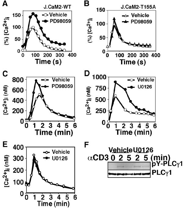
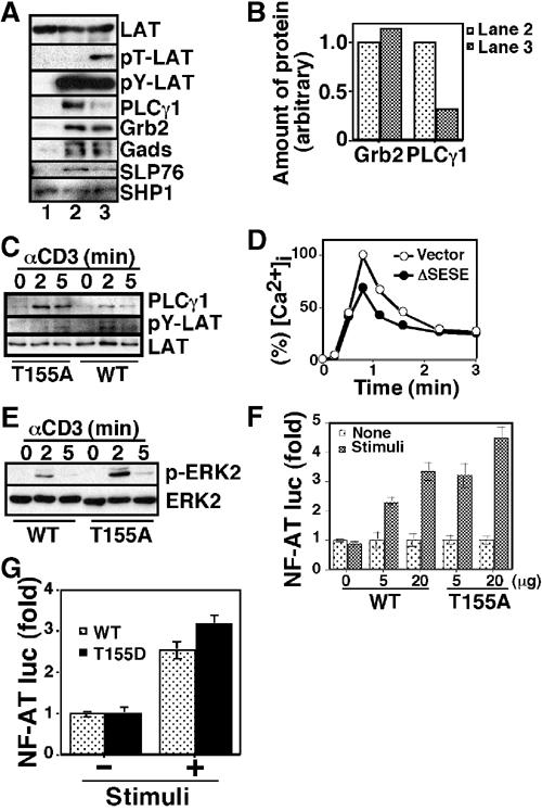
Mitogen-activated protein kinase (MAPK) cascades are involved in a variety of cellular responses including proliferation, differentiation, and apoptosis. We have developed an expression screening method to detect in vivo substrates of MAPKs in mammalian cells, and identified a membrane protein, linker for activation of T cells (LAT), as an MAPK target. LAT, an adapter protein essential for T-cell signaling, is phosphorylated at its Thr 155 by ERK in response to T-cell receptor stimulation. Thr 155 phosphorylation reduces the ability of LAT to recruit PLCgamma1 and SLP76, leading to attenuation of subsequent downstream events such as [Ca2+]i mobilization and activation of the ERK pathway. Our data reveal a new role for MAPKs in a negative feedback loop in T-cell activation via threonine phosphorylation of LAT.


Figures

Figure 1
Figure 1
Cloning of LAT as a putative target of the MAPK pathway. (A) Outline of the screening method. GFP-positive cells were sorted on a FACS Vantage (Becton Dickinson). The resultant clones were subjected to a second screening with pFR-Luc (Stratagene) along with pRL-SV40 (Promega), and assayed for luciferase activity using the Dual-Luc assay system (Promega). (B) Schematic diagram of clone #5–8 and LAT. Potential MAPK phosphorylation sites are indicated. (C) Phosphoamino acid analysis of LAT. 32P-labeled LAT molecules were immunoprecipitated from TCR-stimulated Jurkat cell lysates (human) or AE7 cell lysates (mouse), and subjected to phosphoamino acid analysis. (D) MAPK-dependent activation of clone #5–8. Jurkat cells were transiently transfected with clone #5–8, pFR-Luc, and pRL-SV40 along with pFC-MEKK1 (an expression vector for ΔMEKK1, Stratagene) and/or pSRαMyc-MKP5. Luciferase activities measured in triplicate are expressed as fold increase.
Figure 2
Figure 2
Phosphorylation of LAT at Thr 155 by JNK and ERK. (A) GST-LAT proteins were incubated with active JNK (+) or inactive JNK (−) in the presence of [γ-32P]ATP at 30°C for 30 min. (B) GST-LAT proteins were phosphorylated with active JNK (lanes 2–5) or inactive JNK (lane 1), and subjected to immunoblot with an anti-phospho-Thr-Pro (Cell Signaling) antibody. (C) GST-LAT proteins were phosphorylated with active ERK, and subjected to immunoblot with the anti-phospho-Thr-Pro antibody. (D) GST-LAT or GST-ATF2 proteins were phosphorylated with active (+) or inactive (−) p38, and subjected to immunoblot with the anti-phospho-Thr-Pro antibody (upper) or anti-phospho-ATF2 antibody (lower). (E) LAT proteins were immunoprecipitated from transiently transfected COS7 cells, and subjected to immunoblot with the anti-phospho-Thr-Pro antibody or the antibody against LAT. (F) GFP-fused mouse LAT proteins were immunoprecipitated from transiently transfected Jurkat cells, which are cotransfected with (+) or without (−) ΔSESE, and subjected to immunoblot with the anti-phospho-Thr-Pro antibody or the antibody against GFP.
Figure 3
Figure 3
LAT phosphorylation at T155 during T-cell activation. (A) J.CaM2 cells transfected with WT or T155A mutant LAT were stimulated with anti-CD3ɛ mAb 2Ad2A2 for the indicated times. Immunoprecipitated LAT was subjected to immunoblot with 4G10 or the mAb against phospho-Thr-Pro. (B) Jurkat cells were stimulated with 2Ad2A2 for the indicated times (αCD3) or with pervanadate for 10 min (PV). The lysates were subjected to immunoblot analysis with an mAb against phospho-ERK (Cell Signaling) (p-ERK2) and the anti-ERK2 antibody (ERK2). The lysates were also subjected to immunoprecipitation with anti-JNK1 antibody followed by the kinase assay using ATF2 as a substrate (p-ATF2). Immunoprecipitation of JNK1 was confirmed with immunoblot (JNK). (C) Jurkat cells were stimulated with (+) or without (−) 2Ad2A2 for 3 min in the presence or absence of 50 μM PD98059. Immunoprecipitated LAT was subjected to immunoblot with the mAb against phospho-Thr-Pro (upper panel) or 4G10 (an mAb against phospho-Tyr) (lower panel).
Figure 4
Figure 4
Effects of LAT phosphorylation at T155 on LAT function. (A) The cells were assayed for TCR-induced Ca2+ influx. [Ca2+]i levels were measured on a flow cytometer (right), and calculated as described in Materials and methods (left). (B) J.CaM2 cells stably expressing Myc-tagged LAT were assayed for TCR-induced Ca2+ influx (left). LAT expression levels were confirmed by immunoblot with the anti-LAT antibody and an anti-ZAP70 mAb (Transduction Labs.) (right). It should be noted that wild-type LAT is more expressed compared to T155A mutants. (C) The cells were stimulated with (+) or without (−) 2Ad2A2 for 2 min. ZAP70 and LAT were immunoprecipitated, and subjected to immunoblot with 4G10. (D) Raft fractions were prepared as described in Materials and methods. Existence of LAT was detected with the anti-LAT antibody.
Figure 5
Figure 5
Effect of LAT phosphorylation at T155 on TCR-induced Ca2+ influx. J.CaM2-WT cells (A), J.CaM2-T155A cells (B), human peripheral T cells (C, D), or mouse splenic T cells (E) were assayed for Ca2+ influx. The cells were pretreated with or without the indicated inhibitor (50 μM PD98059 or 10 μM U0126) for 10 min. The data (A, B) are shown as % [Ca2+]i relative to the peak concentration of the vehicle-treated cells (170 nM for J.CaM2-WT; 331 nM for J.CaM2-T155A). (F) J.CaM2-WT cells were stimulated with 2Ad2A2 in the presence or absence of U0126 for the indicated times. The lysates were subjected to immunoprecipitation with an anti-PLCγ1 mAb (Upstate Biotechnology), followed by immunoblot analysis with 4G10 (upper) and the anti-PLCγ1 mAb (lower).
Figure 6
Figure 6
Effect of LAT phosphorylation at T155 on signalsome formation. (A) Jurkat cell lysates were subjected to pull-down assay using mock-treated (lane 1), tyrosine-phosphorylated (lane 2), or tyrosine and threonine doubly phosphorylated (lane 3) GST-LAT molecules. Precipitated GST-LAT proteins and their phosphorylation status were confirmed by immunoblot analysis with the anti-LAT antibody (LAT), the mAb against phospho-Thr-Pro (pT-LAT), and 4G10 (pY-LAT), respectively. Associated proteins were probed with the anti-PLCγ1 mAb, an anti-Grb2 mAb (Transduction Labs.), an anti-Gads antibody (Upstate Biotechnology), an anti-SLP76 antibody (Upstate Biotechnology), and an anti-SHP1 antibody (Upstate Biotechnology). (B) Immunoreactive bands in (A) were quantified, and amounts of PLCγ1 and Grb2 relative to tyrosine-phosphorylated LAT (pY-LAT) were indicated. (C) J.CaM2-WT cells (WT) or J.CaM2-T155A cells (T155A) were stimulated with 2Ad2A2 for indicated times, and then the lysates were subjected to immunoprecipitation with the anti-LAT antibody followed by immunoblot analysis with the anti-PLCγ1 mAb (PLC-γ1) and 4G10 (pY-LAT). (D) Jurkat cells were transiently transfected with an expression vector for human CD8 along with (ΔSESE) or without (vector) an expression vector for ΔSESE. The transfected cells were purified by an anti-human CD8 mAb conjugated with MicroBeads (Miltenyi Biotech GmbH) using an autoMACS (Miltenyi Biotech GmbH), and assayed for Ca2+ influx. The data are shown as % [Ca2+]i relative to the peak concentration of the vector-transfected cells (558 nM). (E) J.CaM2-WT cells (WT) or J.CaM2-T155A cells (T155A) were stimulated with 2Ad2A2 for the indicated times. The lysates were subjected to immunoblot analysis with an mAb against phospho-ERK (upper) and the anti-ERK2 antibody (lower). (F, G) J.CaM2 cells were transiently transfected with NF-AT luc along with the indicated amount of LAT expression vectors, followed by incubation for 40 h. The cells were stimulated with or without 2Ad2A2 in combination with PMA for 7 h, and assayed for luciferase activities.

Similar articles

Cited by

References

    1. Aguado E, Richelme S, Nunez-Cruz S, Miazek A, Mura AM, Richelme M, Guo XJ, Sainty D, He HT, Malissen B, Malissen M (2002) Induction of T helper type 2 immunity by a point mutation in the LAT adaptor. Science 296: 2036–2040 - PubMed
    1. Ahn NG, Seger R, Krebs EG (1992) The mitogen-activated protein kinase activator. Curr Opin Cell Biol 4: 992–999 - PubMed
    1. Alberola-Ila J, Forbush KA, Seger R, Krebs EG, Perlmutter RM (1995) Selective requirement for MAP kinase activation in thymocyte differentiation. Nature 373: 620–623 - PubMed
    1. Alessi DR, Cuenda A, Cohen P, Dudley DT, Saltiel AR (1995) PD 098059 is a specific inhibitor of the activation of mitogen-activated protein kinase kinase in vitro and in vivo. J Biol Chem 270: 27489–27494 - PubMed
    1. Allan LA, Morrice N, Brady S, Magee G, Pathak S, Clarke PR (2003) Inhibition of caspase-9 through phosphorylation at Thr 125 by ERK MAPK. Nat Cell Biol 5: 647–654 - PubMed

Publication types

MeSH terms