Review
. 2004 Aug 15;382(Pt 1):1-11.
doi: 10.1042/BJ20040825.

PTEN function: how normal cells control it and tumour cells lose it

Affiliations
Review

PTEN function: how normal cells control it and tumour cells lose it

Nick R Leslie et al. Biochem J. .

Abstract

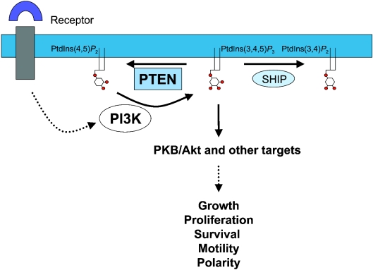
The PTEN (phosphatase and tensin homologue deleted on chromosome 10) tumour suppressor is a PI (phosphoinositide) 3-phosphatase that can inhibit cellular proliferation, survival and growth by inactivating PI 3-kinase-dependent signalling. It also suppresses cellular motility through mechanisms that may be partially independent of phosphatase activity. PTEN is one of the most commonly lost tumour suppressors in human cancer, and its deregulation is also implicated in several other diseases. Here we discuss recent developments in our understanding of how the cellular activity of PTEN is regulated, and the closely related question of how this activity is lost in tumours. Cellular PTEN function appears to be regulated by controlling both the expression of the enzyme and also its activity through mechanisms including oxidation and phosphorylation-based control of non-substrate membrane binding. Therefore mutation of PTEN in tumours disrupts not only the catalytic function of PTEN, but also its regulatory aspects. However, although mutation of PTEN is uncommon in many human tumour types, loss of PTEN expression seems to be more frequent. It is currently unclear how these tumours lose PTEN expression in the absence of mutation, and while some data implicate other potential tumour suppressors and oncogenes in this process, this area seems likely to be a key focus of future research.


Figures

Figure 1
Figure 1. Model for the inhibition of PI 3-kinase signalling by PTEN and SHIP at the plasma membrane
The lipid second messenger PtdIns(3,4,5)P3 is produced from the abundant cellular PI PtdIns(4,5)P2 by the action of class I PI 3-kinase (PI3K) enzymes, which phosphorylate the 3-position of the inositol ring. PI 3-kinases are activated by diverse stimuli, including many that act through transmembrane growth factor receptors. PtdIns(3,4,5)P3 is metabolized by two classes of phosphatases, exemplified by the PI 3-phosphatase, PTEN, which converts PtdIns(3,4,5)P3 back into PtdIns(4,5)P2, and the SHIP 5-phosphatases that convert PtdIns(3,4,5)P3 into PtdIns(3,4)P2. PtdIns(3,4,5)P3 mediates effects on downstream signalling and cellular behaviour through protein targets, such as PKB/Akt, that are able to recognize this lipid and bind it selectively.
Figure 2
Figure 2. The PTEN protein
The 403-amino-acid PTEN protein is represented. The N-terminal phosphatase domain (amino acids 7–185) and the C2 domain (186–351) are both required for enzymic activity. The catalytic cysteine residue (Cys-124) is represented by a large encircled letter C, and Cys-71 by a smaller letter c. These residues form a reversible disulphide bond when the enzyme becomes oxidized. The N-terminal PtdIns(4,5)P2 binding motif is shown at the N-terminal end of the phosphatase domain, although it is uncertain whether in cells this motif responds to PtdIns(4,5)P2, or perhaps another more abundant acidic lipid such as phosphatidylserine. The extreme C-terminal PDZ binding sequence is also shown, and although it is represented as a small region, the extent of further sequences required for optimal specificity and affinity of binding is not known. The phosphorylation sites in the C-terminal tail are represented by a circled letter P.
Figure 3
Figure 3. Model for regulation of PTEN activity by phosphorylation
A model for the regulation of PTEN activity has been developed, largely from the work of Vazquez et al. [43,46] and Das et al. [51]. Phosphorylated residues in the C-terminal tail of PTEN interact with basic regions of the phosphatase (Phos) and C2 domains, causing the long unstructured C-terminal tail to mask the basic membrane-binding surface of PTEN. This causes PTEN to remain in the cytosol, away from its membrane-located substrate, PtdIns(3,4,5)P3. Dephosphorylation of these residues releases the C-terminal tail, allowing the electrostatic interaction of the phosphatase and C2 domains of PTEN with acidic membranes. This also enhances protein–protein interactions mediated by the C-terminal tail, particularly through the PDZ binding site. It is important to note that dephosphorylation of PTEN also enhances its proteolytic degradation.

References

    1. Vanhaesebroeck B., Leevers S. J., Ahmadi K., Timms J., Katso R., Driscoll P. C., Woscholski R., Parker P. J., Waterfield M. D. Synthesis and function of 3-phosphorylated inositol lipids. Annu. Rev. Biochem. 2001;70:535–602. - PubMed
    1. Foster F. M., Traer C. J., Abraham S. M., Fry M. J. The phosphoinositide (PI) 3-kinase family. J. Cell Sci. 2003;116:3037–3040. - PubMed
    1. Katso R., Okkenhaug K., Ahmadi K., White S., Timms J., Waterfield M. D. Cellular function of phosphoinositide 3-kinases: implications for development, homeostasis, and cancer. Annu. Rev. Cell Dev. Biol. 2001;17:615–675. - PubMed
    1. Cantley L. C. The phosphoinositide 3-kinase pathway. Science. 2002;296:1655–1657. - PubMed
    1. Cantrell D. A. Phosphoinositide 3-kinase signalling pathways. J. Cell Sci. 2001;114:1439–1445. - PubMed

Publication types

Substances