

The angiostatic activity of interferon-inducible protein-10/CXCL10 in human melanoma depends on binding to CXCR3 but not to glycosaminoglycan
- PMID: 15194051
- PMCID: PMC2668261
- DOI: 10.1016/j.ymthe.2004.01.010
The angiostatic activity of interferon-inducible protein-10/CXCL10 in human melanoma depends on binding to CXCR3 but not to glycosaminoglycan
Abstract
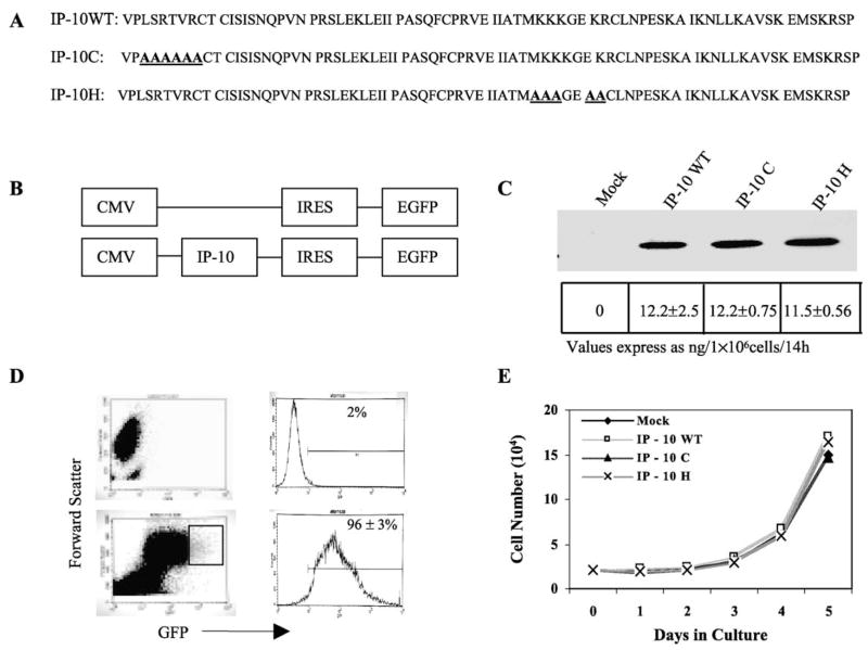
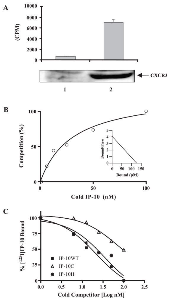
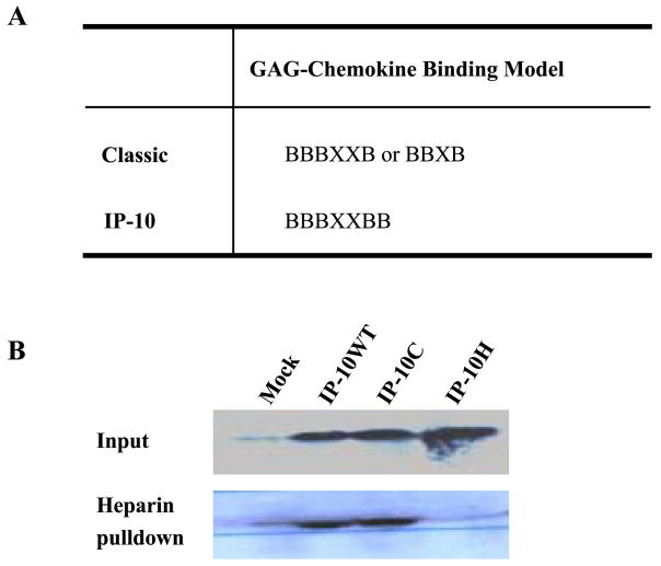
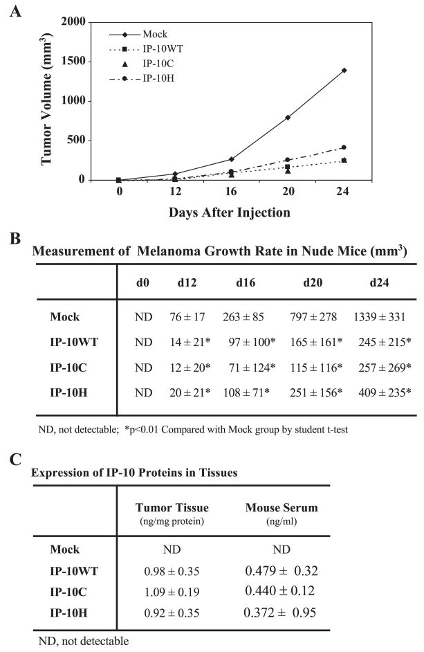
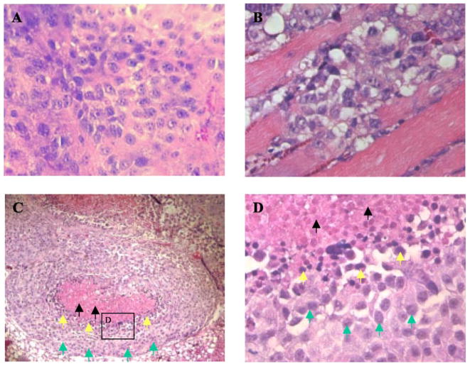
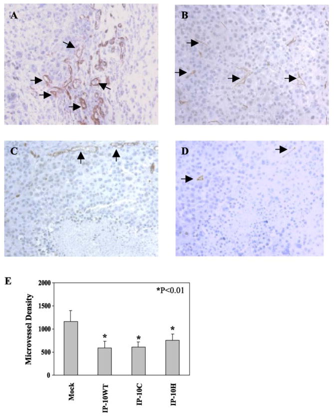
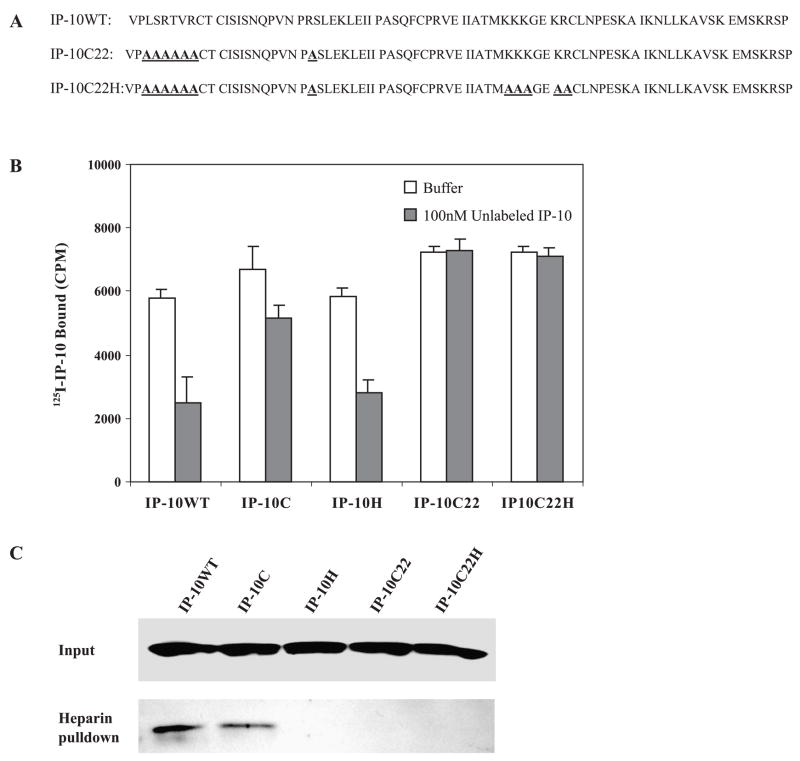
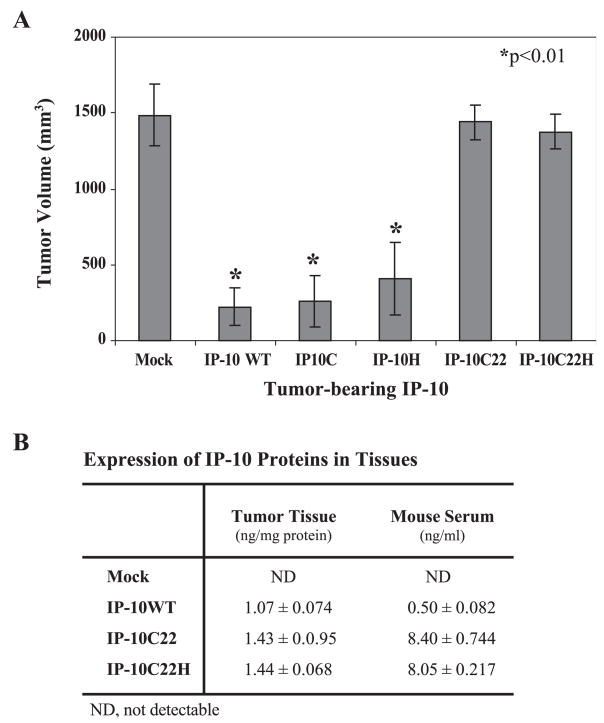
Human interferon-inducible protein 10 (IP-10; HGMW-approved gene symbol CXCL10) is an ELR(-) CXC chemokine that contains binding domains for both the chemokine receptor CXCR3 and glycosaminoglycans. IP-10 has been recently demonstrated to be a potent angiostatic protein in vivo. Whether IP-10 exerts its angiostatic function through binding to CXCR3, glycosaminoglycans, or both, is not clear. To clarify this issue, we created expression constructs for mutants of IP-10 that exhibit partial (IP-10C) or total (IP-10C22) loss of binding to CXCR3 or loss of binding to glycosaminoglycans (IP-10H and IP-10C22H). The A375 human melanoma cell line was transfected with these expression vectors, and stable clones were selected and inoculated subcutaneously into nude mice. As expected, tumor cells secreting wild-type IP-10 showed remarkable reduction in tumor growth compared to control vector-transfected tumor cells. Surprisingly, mutation of IP-10 resulting in partial loss of receptor binding (IP-10C), or loss of GAG binding (IP-10H), did not significantly alter the ability to inhibit tumor growth. This tumor growth inhibition was associated with a reduction in microvessel density, leading to the observed increase in both tumor cell apoptosis and necrosis. In contrast, expression of the IP-10C22 mutant failed to inhibit melanoma tumor growth. These data suggest that CXCR3 receptor binding, but not glycosaminoglycan binding, is essential for the tumor angiostatic activity of IP-10. We conclude that the arginine 22 amino acid residue of IP-10 is essential for both CXCR3 binding and angiostasis.
Figures
Similar articles
-
A functional IFN-gamma-inducible protein-10/CXCL10-specific receptor expressed by epithelial and endothelial cells that is neither CXCR3 nor glycosaminoglycan.J Immunol. 2001 Dec 1;167(11):6576-82. doi: 10.4049/jimmunol.167.11.6576. J Immunol. 2001. PMID: 11714827
-
CXCR3 and heparin binding sites of the chemokine IP-10 (CXCL10).J Biol Chem. 2003 May 9;278(19):17066-74. doi: 10.1074/jbc.M212077200. Epub 2003 Feb 5. J Biol Chem. 2003. PMID: 12571234
-
An alternatively spliced variant of CXCR3 mediates the inhibition of endothelial cell growth induced by IP-10, Mig, and I-TAC, and acts as functional receptor for platelet factor 4.J Exp Med. 2003 Jun 2;197(11):1537-49. doi: 10.1084/jem.20021897. J Exp Med. 2003. PMID: 12782716 Free PMC article.
-
CXCR3, a double-edged sword in tumor progression and angiogenesis.Biochim Biophys Acta. 2013 Dec;1836(2):287-95. doi: 10.1016/j.bbcan.2013.08.002. Epub 2013 Aug 27. Biochim Biophys Acta. 2013. PMID: 23994549 Review.
-
CXC chemokines in angiogenesis relevant to chronic fibroproliferation.Curr Drug Targets Inflamm Allergy. 2005 Feb;4(1):23-6. doi: 10.2174/1568010053622902. Curr Drug Targets Inflamm Allergy. 2005. PMID: 15720231 Review.
Cited by
-
The role of CXC chemokines and their receptors in the progression and treatment of tumors.J Mol Histol. 2012 Dec;43(6):699-713. doi: 10.1007/s10735-012-9435-x. Epub 2012 Jun 30. J Mol Histol. 2012. PMID: 22752457 Review.
-
The response of VEGF-stimulated endothelial cells to angiostatic molecules is substrate-dependent.BMC Cell Biol. 2005 Oct 31;6:38. doi: 10.1186/1471-2121-6-38. BMC Cell Biol. 2005. PMID: 16262896 Free PMC article.
-
Overview of the Mechanisms that May Contribute to the Non-Redundant Activities of Interferon-Inducible CXC Chemokine Receptor 3 Ligands.Front Immunol. 2018 Jan 15;8:1970. doi: 10.3389/fimmu.2017.01970. eCollection 2017. Front Immunol. 2018. PMID: 29379506 Free PMC article. Review.
-
Chemokines as mediators of tumor angiogenesis and neovascularization.Exp Cell Res. 2011 Mar 10;317(5):685-90. doi: 10.1016/j.yexcr.2010.10.020. Epub 2010 Oct 30. Exp Cell Res. 2011. PMID: 21040721 Free PMC article. Review.
-
Human innate lymphoid cell activation by adenoviruses is modified by host defense proteins and neutralizing antibodies.Front Immunol. 2022 Oct 5;13:975910. doi: 10.3389/fimmu.2022.975910. eCollection 2022. Front Immunol. 2022. PMID: 36275713 Free PMC article.
References
-
- Luster AD, Unkeless JC, Ravetch JV. Gamma-interferon transcriptionally regulates an early-response gene containing homology to platelet proteins. Nature. 1985;315:672 – 676. - PubMed
-
- Zaks-Zilberman M, Zaks TZ, Vogel SN. Induction of proinflammatory and chemokine genes by lipopolysaccharide and paclitaxel (Taxol) in murine and human breast cancer cell lines. Cytokine. 2001;15:156 – 165. - PubMed
Publication types
MeSH terms
Substances
Grants and funding
LinkOut - more resources
Full Text Sources
Other Literature Sources
Medical
Research Materials
