

Macrophage specificity of three anti-CD68 monoclonal antibodies (KP1, EBM11, and PGM1) widely used for immunohistochemistry and flow cytometry
- PMID: 15194571
- PMCID: PMC1755048
- DOI: 10.1136/ard.2003.013029
Macrophage specificity of three anti-CD68 monoclonal antibodies (KP1, EBM11, and PGM1) widely used for immunohistochemistry and flow cytometry
Abstract
Objectives: To investigate the specificity of three anti-CD68 monoclonal antibodies (mAbs) for macrophages (Mphi) in immunohistochemistry (IHC) and flow cytometry (FACS).
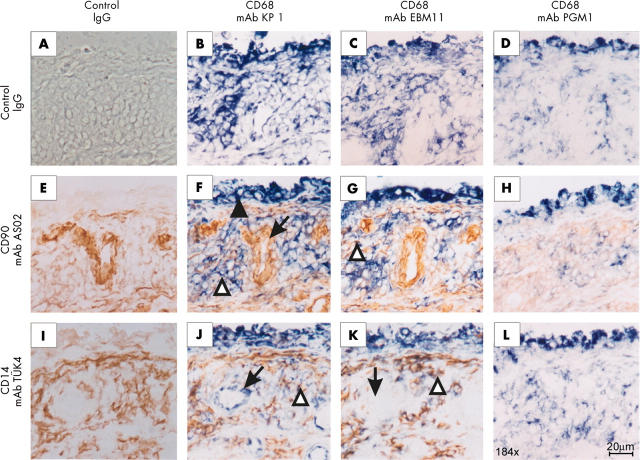
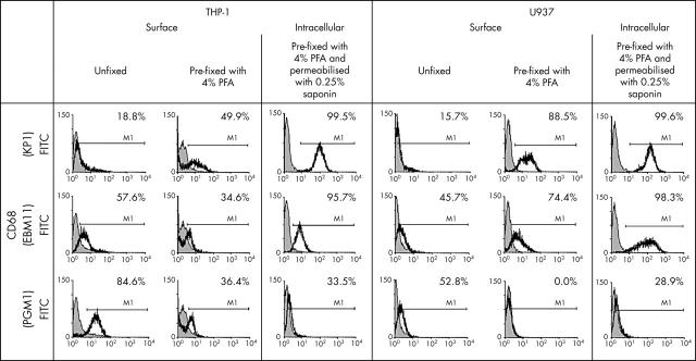
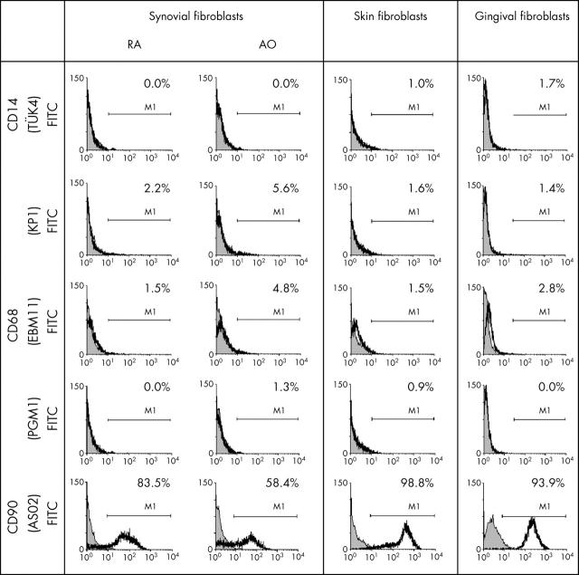
Methods: IHC was performed on cryostat sections of rheumatoid arthritis (RA) and osteoarthritis (OA) synovial membranes using the anti-CD68 mAbs KP1, EBM11, and PGM1, and the fibroblast (FB) markers CD90 and prolyl 4-hydroxylase. Expression of CD68 was also analysed by FACS on the monocytic cell lines THP-1 and U937, as well as on synovial fibroblasts (SFB), skin FB, and gingival FB (both surface and intracellular staining).
Results: In IHC, there was an overlap between CD68 (mAbs KP1 and EBM11) and the FB markers CD90/prolyl 4-hydroxylase in the lining layer, diffuse infiltrates, and stroma of RA and OA synovial membranes. In FACS analysis of THP-1 and U937 cells, the percentage of cells positive for the anti-CD68 mAbs KP1 and EBM11 progressively increased from surface staining of unfixed cells, to surface staining of pre-fixed cells, to intracellular staining of the cells. Upon intracellular FACS of different FB, nearly all cells were positive for KP1 and EBM11, but only a small percentage for PGM1. In surface staining FACS, a small percentage of FB were positive for all three anti-CD68 mAbs.
Conclusion: An overlap between CD68 (mAbs KP1 or EBM11) and the FB markers CD90 or prolyl 4-hydroxylase may prevent unequivocal identification of Mphi in synovial tissue by IHC or in monocytic cells and FB upon intracellular FACS. This may be due to sharing of common markers by completely different cell lineages.
Figures

Comment in
-
CD68 is not a macrophage-specific antigen.Ann Rheum Dis. 2005 Feb;64(2):342-3; author reply 343-4. Ann Rheum Dis. 2005. PMID: 15647451 Free PMC article. No abstract available.
Similar articles
-
Inhomogeneity of immune cell composition in the synovial sublining: linear mixed modelling indicates differences in distribution and spatial decline of CD68+ macrophages in osteoarthritis and rheumatoid arthritis.Arthritis Res Ther. 2016 Jul 16;18:170. doi: 10.1186/s13075-016-1057-3. Arthritis Res Ther. 2016. PMID: 27424032 Free PMC article.
-
KP1 (CD68)-positive large cell lymphomas: a histopathologic and immunophenotypic characterization of 12 cases.Hum Pathol. 1993 Aug;24(8):886-96. doi: 10.1016/0046-8177(93)90139-8. Hum Pathol. 1993. PMID: 7690736
-
PG-M1: a new monoclonal antibody directed against a fixative-resistant epitope on the macrophage-restricted form of the CD68 molecule.Am J Pathol. 1993 May;142(5):1359-72. Am J Pathol. 1993. PMID: 7684194 Free PMC article.
-
Macrophages expressing the scavenger receptor CD163: a link between immune alterations of the gut and synovial inflammation in spondyloarthropathy.J Pathol. 2002 Mar;196(3):343-50. doi: 10.1002/path.1044. J Pathol. 2002. PMID: 11857499
-
[Macrophages in rheumatoid synovial membrane: an update].Rev Rhum Ed Fr. 1993 Oct;60(9):568-79. Rev Rhum Ed Fr. 1993. PMID: 8012331 Review. French.
Cited by
-
Investigation of local stimulation effects of embedding PGLA at Zusanli (ST36) acupoint in rats based on TRPV2 and TRPV4 ion channels.Front Neurosci. 2024 Oct 9;18:1469142. doi: 10.3389/fnins.2024.1469142. eCollection 2024. Front Neurosci. 2024. PMID: 39445077 Free PMC article.
-
Is Synovial Macrophage Activation the Inflammatory Link Between Obesity and Osteoarthritis?Curr Rheumatol Rep. 2016 Sep;18(9):57. doi: 10.1007/s11926-016-0605-9. Curr Rheumatol Rep. 2016. PMID: 27422277 Review.
-
The Mac Is Back: The Role of Macrophages in Human Healthy and Complicated Pregnancies.Int J Mol Sci. 2023 Mar 10;24(6):5300. doi: 10.3390/ijms24065300. Int J Mol Sci. 2023. PMID: 36982375 Free PMC article. Review.
-
Regulation of inflammation-associated olfactory neuronal death and regeneration by the type II tumor necrosis factor receptor.Int Forum Allergy Rhinol. 2013 Sep;3(9):740-7. doi: 10.1002/alr.21187. Epub 2013 Jun 3. Int Forum Allergy Rhinol. 2013. PMID: 23733314 Free PMC article.
-
Adipocyte dysfunction in a mouse model of polycystic ovary syndrome (PCOS): evidence of adipocyte hypertrophy and tissue-specific inflammation.PLoS One. 2012;7(10):e48643. doi: 10.1371/journal.pone.0048643. Epub 2012 Oct 31. PLoS One. 2012. PMID: 23119079 Free PMC article.
References
Publication types
MeSH terms
Substances
LinkOut - more resources
Full Text Sources
Other Literature Sources
Medical
Molecular Biology Databases
Miscellaneous
