

Immunohistochemistry of normal human knee synovium: a quantitative study
- PMID: 15194572
- PMCID: PMC1755068
- DOI: 10.1136/ard.2003.013383
Immunohistochemistry of normal human knee synovium: a quantitative study
Abstract
Objective: To describe the immunohistochemical characteristics of knee synovium from normal healthy subjects.
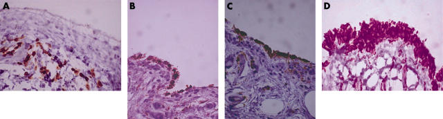
Methods: 12 healthy subjects underwent needle biopsy of knee synovium. Using antibodies directed against CD3, CD4, CD8, L26, Kp-1,and HLA-DR, detailed quantitative immunohistochemical analysis of various cell subpopulations was undertaken.
Results: The mean (SD) age of the subjects was 37 (9) years (five male, seven female). All had a negative history for arthritis, no knee pain, and a totally normal joint examination except for the presence of retropatellar crepitus in five. For technical reasons staining for all immunohistochemical markers could not be achieved in all subjects. CD3+ T lymphocytes were seen in nine of 10 subjects, either diffusely or, more commonly, in perivascular areas. CD4+ cells were seen in synovium in three of seven subjects and CD8+ cells in six of eight subjects, in almost equal numbers (CD4:CD8, 1.1:1). L26+ B lymphocytes were not seen in any biopsy. Kp1+ macrophages were found in 10 of 10 subjects, predominantly in surface lining cells, and in small numbers in diffuse and perivascular locations. HLA-DR+ cells were seen in 10 of 10 subjects, predominantly in surface lining cells and diffusely, but a few were seen perivascularly.
Conclusions: Synovium from apparently normal subjects contained a wide range of different cell subpopulations but no B cells. The significance of these immune cells in normal synovium is unclear. A better understanding of their role in normal synovium may be important in analysing the transition to synovitis.
Figures
Comment in
-
Immunohistochemistry of normal synovium.Ann Rheum Dis. 2004 Nov;63(11):1532-3; author reply 1533. Ann Rheum Dis. 2004. PMID: 15479918 Free PMC article. No abstract available.
References
Publication types
MeSH terms
Substances
LinkOut - more resources
Full Text Sources
Other Literature Sources
Research Materials
