Comparative Study
. 2004 Aug 15;559(Pt 1):103-20.
doi: 10.1113/jphysiol.2004.063347. Epub 2004 Jun 11.

Heterogeneous expression of repolarizing, voltage-gated K+ currents in adult mouse ventricles

Affiliations
Comparative Study

Heterogeneous expression of repolarizing, voltage-gated K+ currents in adult mouse ventricles

Sylvain Brunet et al. J Physiol. .

Erratum in

  • J Physiol. 2004 Sep 15;559(Pt 3):985

Abstract

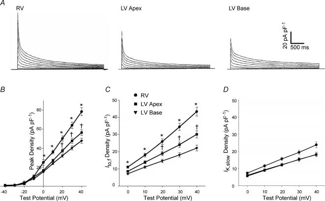
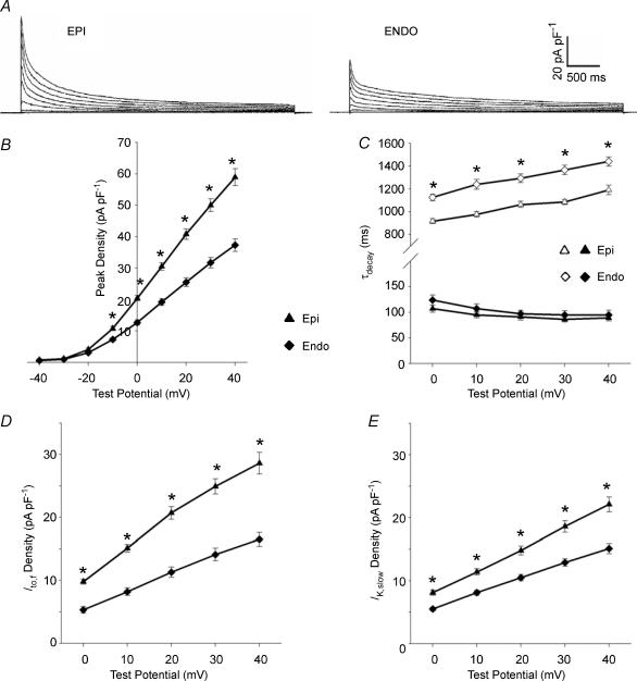
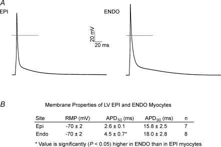
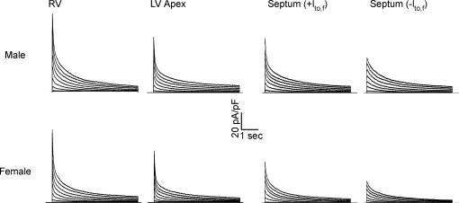
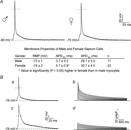
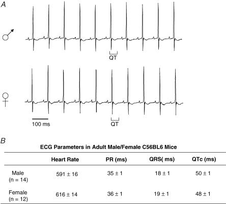
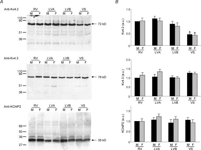
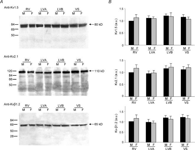
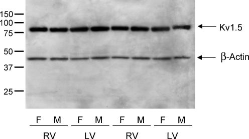
Previous studies have documented the expression of four kinetically distinct voltage-gated K(+) (Kv) currents, I(to,f), I(to,s), I(K,slow) and I(ss), in mouse ventricular myocytes and demonstrated that I(to,f) and I(to,s) are differentially expressed in the left ventricular apex and the interventricular septum. The experiments here were undertaken to test the hypothesis that there are further regional differences in the expression of Kv currents or the Kv subunits (Kv4.2, Kv4.3, KChIP2, Kv1.5, Kv2.1) encoding these currents in adult male and female (C57BL6) mouse ventricles. Whole-cell voltage-clamp recordings revealed that mean (+/-s.e.m.) peak outward K(+) current and I(to,f) densities are significantly (P < 0.001) higher in cells isolated from the right (RV) than the left (LV) ventricles. Within the LV, peak outward K(+) current and I(to,f) densities are significantly (P < 0.05) higher in cells from the apex than the base. In addition, I(to,f) and I(K,slow) densities are lower in cells isolated from the endocardial (Endo) than the epicardial (Epi) surface of the LV wall. Importantly, similar to LV apex cells, I(to,s) is not detected in RV, LV base, LV Epi or LV Endo myocytes. No measurable differences in K(+) current densities or properties are evident in RV or LV cells from adult male and female mice, although I(to,f), I(to,s), I(K,slow) and I(ss) densities are significantly (P < 0.01) higher, and action potential durations at 50% (APD(50)) are significantly (P < 0.05) shorter in male septum cells. Western blot analysis revealed that the expression levels of Kv4.2, Kv4.3, KChIP2, Kv1.5 and Kv2.1 are similar in male and female ventricles. In addition, consistent with the similarities in repolarizing Kv current densities, no measurable differences in ECG parameters, including corrected QT (QT(c)) intervals, are detected in telemetric recordings from adult male and female (C57BL6) mice.


Figures

Figure 1
Figure 1. Peak outward K+ current and Ito,f densities are higher in adult mouse RV than in LV apex or base myocytes
A, depolarization-activated outward K+ currents, evoked during 4.5 depolarizing voltage steps to potentials between −40 and +40 mV from a HP of −70 mV, were recorded and analysed as described in Methods. B, mean ± s.e.m. peak current densities are significantly (*P < 0.001) higher in RV (n = 26) than in either LV apex (n = 25) or LV base (n = 26) myocytes. In addition, mean ± s.e.m. peak current densities are significantly (+P < 0.05) higher in LV apex (n = 25) than in LV base (n = 26) cells. Analysis of the decay phases of the currents revealed that mean ± s.e.m. Ito,f density (C) is significantly (*P < 0.001) higher in RV (n = 26) than in LV apex (n = 25) or LV base (n = 26) myocytes, and that mean Ito,f density is higher (†P < 0.01) in LV apex than in LV base cells. IK,slow densities in RV, LV apex and LV base myocytes (D), in contrast, are not significantly different.
Figure 2
Figure 2. Peak outward K+ current and Ito,f and IK,slow densities are higher in LV Epi than Endo myocytes
A, outward K+ currents in LV Epi and Endo myocytes were recorded and analysed as described in the legend to Fig. 1. B, mean ± s.e.m. peak outward current densities are significantly (*P < 0.001) higher in LV Epi (n = 20) than LV Endo (n = 27), myocytes. C, the τdecay values for Ito,f (filled symbols) in LV Epi and Endo myocytes are indistinguishable, whereas the τdecay values for IK,slow (open symbols) are significantly (*P < 0.001) larger in LV Endo myocytes. Mean ± s.e.m. Ito,f (D) and IK,slow (E) densities are significantly (*P < 0.001) higher in LV Epi than LV Endo cells.
Figure 3
Figure 3. Action potential waveforms in adult mouse LV Endo and LV Epi myocytes are similar
A, representative action potential waveforms recorded from LV Epi and LV Endo myocytes are displayed; the dotted lines indicate the 0 mV levels. B, summary of the resting and active membrane properties of LV Epi and Endo cells.
Figure 4
Figure 4. Ventricular outward K+ current waveforms in cells from male and female mice are similar
Representative outward K+ current waveforms, recorded as described in the legend to Fig. 1, in ventricular myocytes isolated from the RV, LV apex and septum of adult female and male (C57BL6) mice. Although no significant differences in outward K+ current densities or properties were seen in RV or LV myocytes from male and female mice, K+ current densities are higher in septum cells isolated from adult male (than female) mice (see text and Table 2).
Figure 5
Figure 5. Action potential durations in septum cells are correlated with differences in voltage-gated outward K+ current densities
A, representative action potential waveforms recorded from adult mouse septum cells; the dashed lines indicate the 0 mV levels. A summary of the resting and active membrane properties of adult male and female septum cells is provided below the records. B, action potential durations in septum cells vary with peak outward K+ current densities. Action potentials and depolarization-activated outward K+ currents were recorded from isolated (adult female) septum cells as described in the legends to Figs 3 and 1, respectively; the voltage- and current-clamp recordings in a and b, and c and d were obtained from the same cell. Similar results were obtained in experiments on seven cells.
Figure 6
Figure 6. QTc intervals in adult (C57BL6) male and female animals are indistinguishable
Telemetric ECG recordings were obtained from adult male and female C57BL6 animals as described in Methods. A, telemetric ECG recordings from representative male and female animals are displayed; QT intervals are indicated below the records. B, analyses of ECG recordings revealed no significant differences in any ECG parameters in adult male (n = 14) and female (n = 12) C57BL6 mice. Mean ± s.e.m. heart rate, PR QRS and QTc intervals are indistinguishable in male and female C57BL6 animals.
Figure 7
Figure 7. Expression of voltage-gated K+ channel subunits contributing to Ito,f in adult mouse ventricles
A, ventricular homogenates were prepared from the RV, LV apex, LV base and septum of adult male and female mice, fractionated and immunoblotted with the anti-Kv4.2, anti-Kv4.3, or anti-KChIP2 antibody as described in Methods. Representative Western blots with-anti-Kv4.2 (top), anti-Kv4.3 (middle) and anti-KChIP2 (bottom) are presented in A. Films from individual experiments were scanned directly into a Molecular Dynamics densitometer using Image Quant. The density of the Kv4.2, Kv4.3 or KChIP2 bands in each lane were measured and normalized to the density of the male RV sample on the same blot. Mean ± s.e.m. values from six separate determinations are plotted in B. As reported previously (Guo et al. 2002), Kv4.2 expression is significantly (P < 0.001) lower in the interventricular septum than in the RV or LV and parallels the regional differences in Ito,f density. The expression levels of Kv4.2, Kv4.3 and KChIP2, however, are similar in adult male and female mouse right and left ventricles and in the interventricular septum.
Figure 8
Figure 8. Expression of voltage-gated K+ channel subunits contributing to IK,slow in adult mouse ventricles
A, ventricular homogenates were prepared from the RV, LV apex, LV base and septum of adult male and female mice, fractionated and immunoblotted with the anti-Kv1.5, anti-Kv2.1 or anti-Kvβ1.2 antibody as described in Methods. Representative Western blots with the anti-Kv1.5 (top), anti-Kv2.1 (middle) and anti-Kvβ1.2 (bottom) antibodies are presented in A. Films were scanned as described in the legend to Fig. 7, and the densities of the individual bands were normalized to the density of the male RV sample on the same film. Mean ± s.e.m. values from six separate determinations are plotted in B. The expression levels of Kv1.5, Kv2.1 and Kvβ1.2 are similar in adult male and female mouse RV, LV apex, LV base and interventricular septum.
Figure 9
Figure 9. Expression of Kv1.5, which encodes IK,slow1, in adult male and female CD-1 mouse ventricles
Ventricular homogenates were prepared from the RV and LV of adult male (n = 6) and female (n = 6) CD-1 mice, fractionated and immunoblotted with the anti-Kv1.5 and anti-β-actin antibodies as described in Methods. Representative data from two male and two female RV and LV are presented. Films were scanned as described in the legend to Fig. 7, and no significant differences in Kv1.5 expression were evident in the RV or LV samples from the male and the female CD-1 animals (see text).

Similar articles

Cited by

References

    1. An WF, Bowlby MR, Betty M, Cao J, Li H-P, Mendoza G, Hinson JW, Mattsson KI, Strassle BW, Trimmer JS, Rhodes KJ. Modulation of A-type potassium channels by a family of calcium sensors. Nature. 2000;403:553–556. - PubMed
    1. Antzelevitch C, Dumaine R. Electrical heterogeneity in the heart: Physiological, pharmacological, and clinical implications. In: Page E, Fozzard HA, Solaro RJ, editors. The Handbook of Physiology, section 2, The Cardiovascular System, The Heart. Vol. 1. New York: Oxford University Press; 2002. pp. 654–692.
    1. Anumonwo J, Tallini YN, Vetter FJ, Jalife J. Action potential characteristics and arrhythmogenic properties of the cardiac conduction system of the murine heart. Circ Res. 2001;89:329–335. - PubMed
    1. Baker LC, London B, Choi BR, Koren G, Salama G. Enhanced dispersion of repolarization and refractoriness in transgenic mouse hearts promotes reentrant ventricular tachycardia. Circ Res. 2000;86:396–407. - PubMed
    1. Barry DM, Xu H, Schuessler RB, Nerbonne JM. Functional knockout of the transient outward current, long–QT syndrome, and cardiac remodeling in mice expressing a dominant- negative Kv4 alpha subunit. Circ Res. 1998;83:560–567. - PubMed

Publication types

Substances

LinkOut - more resources