

Review
doi: 10.1128/JVI.78.13.6709-6714.2004.
Pathways of cell infection by parvoviruses and adeno-associated viruses
Affiliations
- PMID: 15194745
- PMCID: PMC421635
- DOI: 10.1128/JVI.78.13.6709-6714.2004
Item in Clipboard
Review
Pathways of cell infection by parvoviruses and adeno-associated viruses
J Virol.
2004 Jul.
No abstract available
Figures
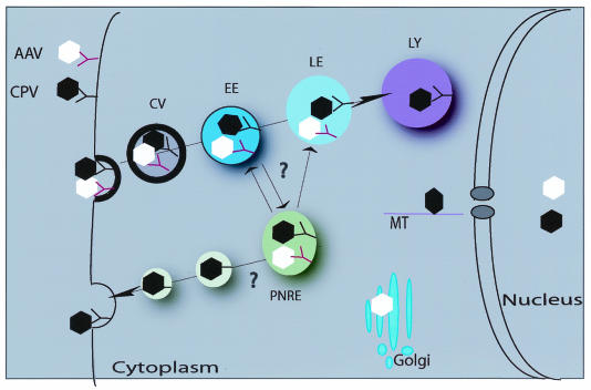
A schematic representation of the entry mechanisms utilized by parvoviruses within the host cell. Cell entry of autonomous parvovirus CPV and AAV is shown. After binding to their cell surface receptors, both viruses are internalized into clathrin-coated vesicles (CV), followed by transport to early (EE), late (LE), or perinuclear recycling endosomes (PNRE). Later in entry, in the case of AAV, capsids are found in Golgi compartments, whereas CPV can be found in lysosomes (LY). The site of the capsid escape from endocytic vesicles into the cytosol is still unclear. CPVs make use of microtubules (MT) during the traffic through the cytosol toward the nucleus. Viral capsids are able to enter the nucleus in intact form without apparent deformation.
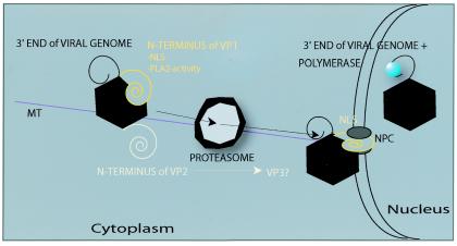
Intracellular trafficking and modifications of autonomous parvovirus capsids. Once inside the cell, capsids undergo modifications, exposing on their surfaces not only the N-terminal end of the VP1 capsid protein but also the N-terminal sequence of the VP2 capsid protein and 3′ end of the viral genome. In the cytosol, capsids are also affected by the activity of proteasomes, possibly causing the intracellular formation of the VP3 capsid protein. Later in entry, viral capsids are imported into the nucleus through the NPC with the help of the NLS. MT, microtubules.
Similar articles
-
The expanding range of parvoviruses which infect humans.Rev Med Virol. 2010 Jul;20(4):231-44. doi: 10.1002/rmv.648. Rev Med Virol. 2010. PMID: 20586082 Review.
-
Parvovirus host range, cell tropism and evolution.Curr Opin Microbiol. 2003 Aug;6(4):392-8. doi: 10.1016/s1369-5274(03)00083-3. Curr Opin Microbiol. 2003. PMID: 12941411 Review.
-
Evolutionary relationships among parvoviruses: virus-host coevolution among autonomous primate parvoviruses and links between adeno-associated and avian parvoviruses.J Virol. 2001 Mar;75(6):2729-40. doi: 10.1128/JVI.75.6.2729-2740.2001. J Virol. 2001. PMID: 11222696 Free PMC article.
-
Emerging Human Parvoviruses: The Rocky Road to Fame.Annu Rev Virol. 2019 Sep 29;6(1):71-91. doi: 10.1146/annurev-virology-092818-015803. Epub 2019 Jul 5. Annu Rev Virol. 2019. PMID: 31283445 Review.
-
Rodent parvovirus infections.Lab Anim Sci. 1996 Aug;46(4):370-80. Lab Anim Sci. 1996. PMID: 8872986 Review.
Cited by
-
Enterovirus 70 binds to different glycoconjugates containing alpha2,3-linked sialic acid on different cell lines.J Virol. 2005 Jun;79(11):7087-94. doi: 10.1128/JVI.79.11.7087-7094.2005. J Virol. 2005. PMID: 15890948 Free PMC article.
-
Recombinant adeno-associated viral vectors are deficient in provoking a DNA damage response.J Virol. 2008 Aug;82(15):7379-87. doi: 10.1128/JVI.00358-08. Epub 2008 May 7. J Virol. 2008. PMID: 18463154 Free PMC article.
-
Visualization of the externalized VP2 N termini of infectious human parvovirus B19.J Virol. 2008 Aug;82(15):7306-12. doi: 10.1128/JVI.00512-08. Epub 2008 May 28. J Virol. 2008. PMID: 18508892 Free PMC article.
-
Virulent variants emerging in mice infected with the apathogenic prototype strain of the parvovirus minute virus of mice exhibit a capsid with low avidity for a primary receptor.J Virol. 2005 Sep;79(17):11280-90. doi: 10.1128/JVI.79.17.11280-11290.2005. J Virol. 2005. PMID: 16103180 Free PMC article.
-
Adeno-associated virus-2 (AAV-2) causes trophoblast dysfunction, and placental AAV-2 infection is associated with preeclampsia.Am J Pathol. 2006 Jun;168(6):1951-9. doi: 10.2353/ajpath.2006.050781. Am J Pathol. 2006. PMID: 16723710 Free PMC article.
References
-
- Agbandje, M., R. McKenna, M. G. Rossmann, M. L. Strassheim, and C. R. Parrish. 1993. Structure determination of feline panleukopenia virus empty particles. Proteins 16:155-171. - PubMed
-
- Agbandje-McKenna, M., A. L. Llamas-Saiz, F. Wang, P. Tattersall, and M. G. Rossmann. 1998. Functional implications of the structure of the murine parvovirus, minute virus of mice. Structure 6:1369-1381. - PubMed
-
- Barbis, D. P., S.-F. Chang, and C. R. Parrish. 1992. Mutations adjacent to the dimple of canine parvovirus capsid structure affect sialic acid binding. Virology 191:301-308. - PubMed
Publication types
MeSH terms
LinkOut - more resources
Full Text Sources
