. 2004 Jul;78(13):6855-63.
doi: 10.1128/JVI.78.13.6855-6863.2004.

Vaccinia virus G1 protein, a predicted metalloprotease, is essential for morphogenesis of infectious virions but not for cleavage of major core proteins

Affiliations

Vaccinia virus G1 protein, a predicted metalloprotease, is essential for morphogenesis of infectious virions but not for cleavage of major core proteins

Camilo Ansarah-Sobrinho et al. J Virol. 2004 Jul.

Abstract

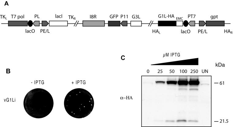
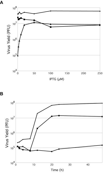
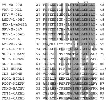
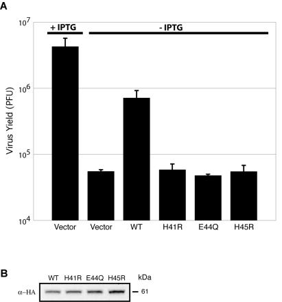
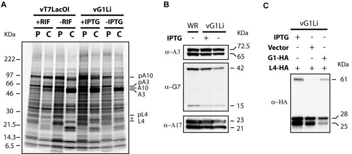
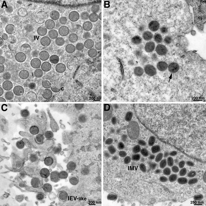
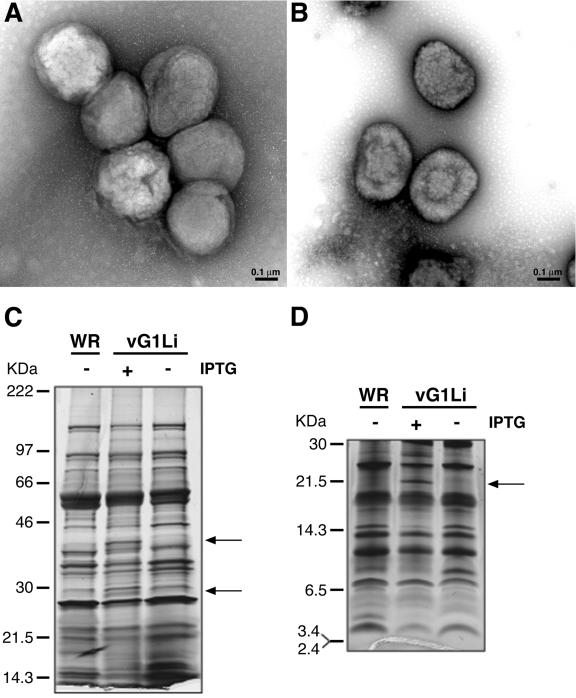
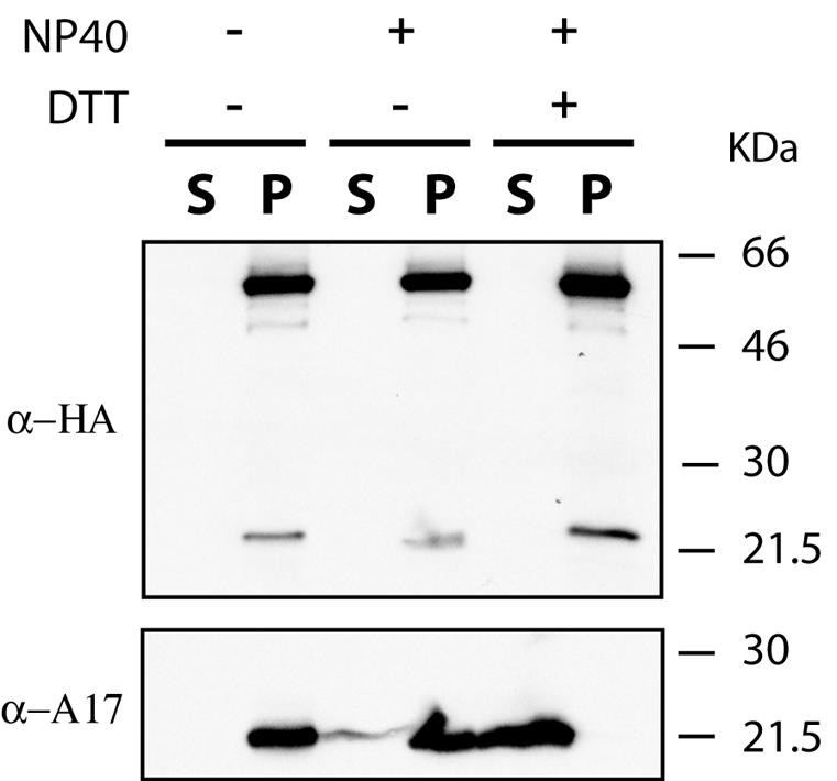
Genes encoding orthologs of the vaccinia virus G1 protein are present in all poxviruses for which sequence information is available, yet neither the role of the protein nor its requirement for virus replication is known. G1 was predicted to be involved in the cleavage of core proteins, based on a transfection study and the presence of an HXXEH motif found in a subset of metallopeptidases. In the present study, we engineered a recombinant vaccinia virus containing a single copy of the G1L gene with a C-terminal epitope tag that is stringently regulated by the Escherichia coli lac repressor. In the absence of inducer, expression of G1 was repressed and virus replication was inhibited. Rescue of infectious virus was achieved by expression of wild-type G1 in trans, but not when the putative protease active site residues histidine-41, glutamate-44, or histidine-45 were mutated. Nevertheless, the synthesis and proteolytic processing of major core and membrane proteins appeared unaffected under nonpermissive conditions, distinguishing the phenotype of the G1L mutant from one in which the gene encoding the I7 protease was repressed. Noninfectious virus particles, assembled in the absence of inducer, did not attain the oval shape or characteristic core structure of mature virions. The polypeptide composition of these particles, however, closely resembled that of wild-type virus. Full-length and shorter forms of the G1 protein were found in the core fraction of virus particles assembled in the presence of inducer, suggesting that G1 is processed by self-cleavage or by another protease.


Figures

FIG. 1.
FIG. 1.
Construction and initial characterization of an inducible G1L mutant. (A) Representation of the genome of vG1Li. Important features include the presence of the bacteriophage T7 RNA polymerase gene (T7 pol) regulated by the VV late P11 promoter (PL) and the E. coli lac operator (lacO); the E. coli lac repressor (lacI) regulated by the VV early-late P7.5 promoter (PE/L); the GFP gene regulated by the late P11 promoter and replacing the original G1L gene; an inducible copy of the G1L gene (G1L-HA) encoding an influenza virus HA epitope tag at the C-terminal end of the protein and regulated by the bacteriophage T7 promoter (PT7); the lacO; and the encephalomyocarditis (EMC) leader. TKL, TKR, HAL, and HAR represent left and right flanking sequences of the VV TK and HA genes. gpt, E. coli guanine phosphoribosyltransferase gene. (B) Effect of IPTG on plaque formation. BS-C-1 cell monolayers were infected with vG1Li in the presence or absence of 50 μM IPTG. After 48 h, plaques were stained and visualized with crystal violet. (C) Effect of IPTG on the expression of G1. Uninfected cells (UN) and cells infected with vG1Li at a multiplicity of infection of 5 in the presence of 0 to 250 μM IPTG were harvested at 18 h after infection. Cell extracts were separated by SDS-PAGE and detected by Western blotting with a MAb to the influenza virus HA epitope tag (α-HA). The masses of marker proteins in kilodaltons are indicated on the right.
FIG. 2.
FIG. 2.
Effect of IPTG on the production of infectious vG1Li. (A) BS-C-1 cells were infected with vT7LacOI (•), vG1L/G1Li (▪), or vG1Li (▴) at a multiplicity of infection of 5 and incubated in the presence of 0 to 250 μM IPTG. At 24 h after infection, virus titers were determined by plaque assay in the presence of 50 μM IPTG. (B) BS-C-1 cells were infected with vT7LacOI (•) or vG1Li in the presence (▪) or absence (▴) of 50 μM IPTG. Cells were harvested at the indicated times after infection, and virus titers were determined as described for panel A.
FIG. 3.
FIG. 3.
Multiple sequence alignment of poxvirus G1L orthologs and HXXEH-containing metallopeptidases. Invariant amino acid residues are shaded gray. The HXXEH motif is indicated by asterisks at the top. The alignment includes two orthopoxvirus sequences, one representative sequence from the other chordopoxvirus genera, and one entomopoxvirus sequence. Numbers indicate first and last amino acids in the alignment. Virus protein names consist of an abbreviation of the virus and gene name as listed in the University of Victoria database (http://www.poxvirus.org/pocs.asp). Nonviral entries consist of abbreviations of gene and organism names. Virus name abbreviations are as follows: VAR-I, variola major virus strain India (GI 420468); VV-WR, Western Reserve strain of VV (GI 335671); FPV-V, fowlpox virus (GI 7271579); LSDV-L, lumpy skin disease virus Neethling vaccine LW 1959 (GI 15149061); MYX-L, myxoma virus strain Lausanne (GI 6523900); SPV-N, sheeppox virus strain NISKHI (GI 21492503); MCV-1, molluscum contagiosum virus subtype 1 (GI 7515386); YLDV, Yaba-like disease virus (GI 12056209); AmEPV, Amsacta moorei entomopoxvirus (GI 9944779). Organism and gene name abbreviations are as follows: PTRA-ECOLI, pitrilysin from E. coli (GI 131573); IDE-DROME, insulin-degrading enzyme from Drosophila melanogaster (GI 124156); IDE-HUMAN, insulin-degrading enzyme from humans (GI 124157); MPPA-HUMAN, mitochondrial processing peptidase alpha subunit from humans (GI 29840846); MPPB-YEAST, mitochondrial processing peptidase beta subunit from Saccharomyces cerevisiae (GI 127290); PQQF-KLEPN, coenzyme pyrroloquinoline quinone biosynthesis protein f from Klebsiella pneumoniae (GI 130803); PQQL-ECOLI, putative coenzyme pyrroloquinoline quinone biosynthesis protein from E. coli (GI 2507259); SDP-EIMBO, sporozoite developmental protein from Eimeria bovis (GI 1173411); YMT1-CAEEL, hypothetical zinc protease f56d2.1 from Caenorhabditis elegans (GI 2507260); YMXG-BACSU, hypothetical zinc protease ymxg from Bacillus subtilis (GI 1176567); YQA4-CAEEL, hypothetical zinc protease c28f5.4 from C. elegans (GI 1730966).
FIG. 4.
FIG. 4.
Complementation of vG1Li with plasmids expressing wild-type G1 but not those expressing mutated G1. (A) BS-C-1 cells were infected with vG1Li at a multiplicity of infection of 3 in the presence or absence of 50 μM IPTG as indicated. After 1 h, cells infected in the absence of IPTG were transfected with 1 μg of vector plasmid or plasmid expressing wild-type (WT) or mutated G1 proteins containing a C-terminal influenza virus HA epitope tag. H41R, histidine-41 mutated to arginine; E44Q, glutamate-44 mutated to glutamine; H45R, histidine-45 mutated to arginine. (B) Lysates from cells infected with vG1Li in the absence of IPTG and transfected with plasmids expressing wild-type or mutated G1-HA were analyzed by Western blotting with MAb to the influenza virus HA epitope (α-HA). The apparent molecular mass in kilodaltons is indicated on the right.
FIG. 5.
FIG. 5.
Synthesis and processing of viral late proteins. (A) BS-C-1 cells were infected with vT7LacOI in the presence or absence of 100 μg of rifampin (RIF) per ml or vG1Li in the presence or absence of 50 μM IPTG. At 8 h after infection, cells were pulse-labeled with a mixture of [35S]methionine and [35S]cysteine for 45 min and either harvested immediately (P) or chased for an additional 16 h with medium containing unlabeled methionine and cysteine (C). Cells were analyzed by SDS-PAGE under reducing conditions and by autoradiography. The protein bands that correspond to the proteolytically processed viral major structural proteins (A3, A10, and L4) and their uncleaved precursors (pA3, pA10, and pL4) are indicated on the right. The asterisk corresponds to the band believed to be GFP. (B) BS-C-1 cells were infected with wild-type VV (WR) or vG1Li in the presence (+) or absence (−) of 50 μM IPTG. At 18 h after infection, cells were harvested and analyzed by Western blotting with antibody to the A3 (α-A3), G7 (α-G7), or A17 (α-A17) protein. Apparent molecular masses in kilodaltons are indicated on the right. (C) BS-C-1 cells were infected with vG1Li in the presence (+) or absence (−) of 50 μM IPTG and transfected with vector plasmid or plasmids expressing G1 (G1-HA) or L4 (L4-HA) protein with an influenza virus HA epitope tag at the C terminus under the control of a VV synthetic strong late promoter. At 18 h after infection, cells were harvested and analyzed by Western blotting with MAb to the influenza virus HA epitope tag (α-HA). Apparent molecular masses in kilodaltons are indicated on the right.
FIG. 6.
FIG. 6.
Electron microscopy of cells infected with vG1Li under nonpermissive conditions. BS-C-1 cells were infected with vG1Li at a multiplicity of infection of 5 in the absence of IPTG (A to C) or with wild-type VV (D). At 24 h after infection, cells were fixed and embedded in Epon, and ultrathin sections were prepared for transmission electron microscopy. (A) Crescents (c) and immature virions (IV), some with nucleoids. (B) Virus particles (arrow) that have progressed beyond the typical IV stage and resemble IMV but are mostly circular with poorly formed cores. (C) IEV-like particles that are wrapped with additional membranes. The images in panels A, B, and C were obtained from different cells in the same thin section. (D) Typical IMV in cells infected with wild-type VV. The intracellular clustering of particles at similar stages of morphogenesis, as seen in each of the panels, is typical.
FIG. 7.
FIG. 7.
Morphology and protein composition of purified vG1Li formed in the presence and absence of IPTG. (A) Electron micrograph of negatively stained virus particles from cells infected with vG1Li in the absence of IPTG and purified by sedimentation through a 36% sucrose cushion followed by a 25-to-40% sucrose gradient. (B) Same experiment as shown in panel A, except that cells were infected with vG1Li in the presence of IPTG. (C) Purified wild-type VV (WR) and vG1Li grown in the presence (+) or absence (−) of IPTG were dissociated with SDS and mercaptoethanol and analyzed by electrophoresis in a 4-to-20% polyacrylamide gel in Tris-glycine buffer. A photograph of a silver-stained gel is shown. Distinctive vG1Li (+ IPTG) bands are indicated by arrows. The molecular masses of the marker proteins are on the left. (D) Same experiment as shown in panel C, but samples were analyzed on a 16% polyacrylamide gel in Tricine buffer.
FIG. 8.
FIG. 8.
Localization of G1 in the virus particle. Sucrose gradient-purified vG1Li, from the preparation shown in Fig. 7B, was incubated in 50 mM Tris-HCl buffer (pH 7.5) alone or with 1% NP-40 or 1% NP-40 plus 50 mM dithiothreitol (DTT). Soluble and insoluble fractions were separated by centrifugation and analyzed by Western blotting with a MAb to the influenza virus HA epitope (α-HA) or polyclonal antibodies to an N-terminal peptide of A17 (α-A17). The molecular masses of the marker proteins are indicated on the right.

Similar articles

Cited by

References

    1. Ansarah-Sobrinho, C., and B. Moss. 2004. Role of the I7 protein in proteolytic processing of vaccinia virus membrane and core components. J. Virol. 78:6335-6343. - PMC - PubMed
    1. Becker, A. B., and R. A. Roth. 1993. Identification of glutamate-169 as the third zinc-binding residue in proteinase III, a member of the family of insulin-degrading enzymes. Biochem. J. 292:137-142. - PMC - PubMed
    1. Becker, A. B., and R. A. Roth. 1992. An unusual active site identified in a family of zinc metalloendopeptidases. Proc. Natl. Acad. Sci. USA 89:3835-3839. - PMC - PubMed
    1. Betakova, T., E. J. Wolffe, and B. Moss. 1999. Regulation of vaccinia virus morphogenesis: phosphorylation of the A14L and A17L membrane proteins and C-terminal truncation of the A17L protein are dependent on the F10L protein kinase. J. Virol. 73:3534-3543. - PMC - PubMed
    1. Byrd, C. M., T. C. Bolken, and D. E. Hruby. 2003. Molecular dissection of the vaccinia virus I7L core protein proteinase. J. Virol. 77:11279-11283. - PMC - PubMed

Publication types

MeSH terms

LinkOut - more resources