. 2004 Jul;78(13):7175-85.
doi: 10.1128/JVI.78.13.7175-7185.2004.

Nuclear sequestration of cellular chaperone and proteasomal machinery during herpes simplex virus type 1 infection

Affiliations

Nuclear sequestration of cellular chaperone and proteasomal machinery during herpes simplex virus type 1 infection

April D Burch et al. J Virol. 2004 Jul.

Abstract

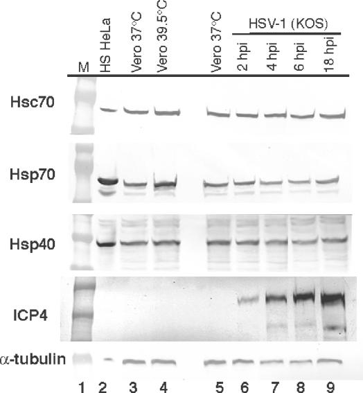
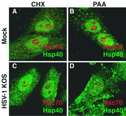
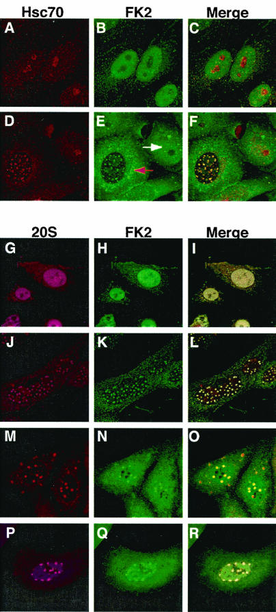
Herpes simplex virus type 1 (HSV-1) encodes a portal protein that forms a large oligomeric structure believed to provide the conduit for DNA entry and exit from the capsid. Chaperone proteins often facilitate the folding and multimerization of such complex structures. In this report, we show that cellular chaperone proteins, components of the 26S proteasome, and ubiquitin-conjugated proteins are sequestered in discrete foci in the nucleus of the infected cell. The immediate-early viral protein ICP0 was shown to be necessary to establish these foci at early times during infection and sufficient to redistribute chaperone molecules in transfected cells. Furthermore, we found that not only is the portal protein, UL6, localized to these sites during infection, but it is also a substrate for ubiquitin modification. Our results suggest that HSV-1 has evolved an elegant mechanism for facilitating protein quality control at specialized foci within the nucleus.


Figures

FIG. 1.
FIG. 1.
Subcellular localization of the cellular chaperone Hsc70/Hsp70 and cochaperone Hsp40 during HSV-1 infection. (A to C, G to I) Uninfected Vero cells cultured for 6 h at 37°C. (D to F, J to L) Vero cells infected with wild-type HSV-1 (strain KOS) for 6 h at 37°C. The staining profile for Hsc70 (red) is shown in panels A and D, the staining profile for Hsp70 (red) is shown in panels G and J, and the staining profile of Hsp40 (green) is shown in panels B, E, H, and K. Merged Hsc70/Hsp40 images are shown in panels C, F, I, and L. White arrows show Hsp40-enriched foci (E and K) that costain with Hsc70 or Hsp70 in the merged image (F and L, respectively).
FIG. 2.
FIG. 2.
Western blot analysis of cellular chaperone molecules (Hsc70, Hsp70, and Hsp40) in HSV-1-infected cell lysates. Lane 1, molecular weight markers; lane 2, heat-shocked HeLa cell lysate; lane 3, uninfected Vero cells cultured at 37°C; lane 4, uninfected Vero cells heat shocked at 39.5°C; lane 5, uninfected Vero cells cultured at 37°C; lanes 6 to 9, HSV-1-infected cell lysates at 2, 4, 6, and 18 h postinfection, respectively. A Western blot of the viral protein ICP4 is shown as a positive control for infection, and a Western blot of α-tubulin was used as a control to ensure that similar amounts of total protein were loaded for all samples.
FIG. 3.
FIG. 3.
Analysis of chaperone localization in infected or uninfected cells treated with CHX or PAA. Merged images of cells stained with antibodies specific to Hsc70 (red) and Hsp40 (green) are shown. (A and B) Uninfected cells treated with CHX or PAA. (C and D) HSV-1-infected cells treated with CHX or PAA.
FIG. 4.
FIG. 4.
Merged images of HSV-1-infected cells stained with antibodies detecting Hsc70 (red) and the viral ICP0 protein (green). Times (in hours) postadsorption are indicated above each panel. (A to C) Infected cells cultured at 37°C; (D to F) infected cells cultured at 39.5°C. In panels C and F, the white arrows indicate ICP0-stained foci that did not costain with Hsc70 and likely represent ND10. The red arrows indicate foci that costained with antibodies recognizing either Hsc70 or ICP0. In panel D, note the colocalization (yellow) between ICP0 and Hsc70 within the nucleolus.
FIG. 5.
FIG. 5.
ICP0 is necessary for chaperone sequestration at early times during infection and sufficient to redistribute the cellular chaperone machinery in transfected cells. Vero cells infected with the 0β mutant (ICP0 mutant) (A to C) at 39.5°C were stained with ICP4 (green) and Hsc70 (red). This temperature was chosen because of the faster kinetics of Hsc70-enriched foci. Thus, at 6 h postexposure to HSV-1, there would have been ample time for Hsc70 focus formation. Staining for the ICP4 protein (green) (A and D) was used as a control for successful infection. As described in the text, in the absence of the ICP0 protein, no redistribution of Hsc70 was observed at this time (B), indicating that ICP0 is necessary for this action early during infection. (D to F) 0β-infected U2OS cells (a human osteosarcoma cell line that naturally complements ICP0 HSV-1 mutants) stained for ICP4 (D) and Hsc70 (E). Merged images are shown in panels C and F. Merged images of HSV-1 (strain KOS)-infected (G) and 0β-infected (H) Vero cells collected at 20 h postinfection are shown as well. (I to K) Subcellular localization of ICP0 (green) and Hsc70 (red) in cells transfected with a plasmid carrying the ICP0 gene. A merged image of the transfected cell is shown in panel K.
FIG. 6.
FIG. 6.
Localization of ubiquitinated proteins and components of the proteasomal machinery during HSV-1 infection. Uninfected (A to C) and HSV-1-infected (D to F) cells stained with antibodies specific to Hsc70 (A and D) and ubiquitinated proteins (FK2) (B and E) are shown. Merged images are shown in panels C and F. In panel E, the white arrow indicates the diffuse nuclear staining of ubiquitinated proteins in an uninfected cell and the red arrow indicates the dramatic redistribution of ubiquitinated proteins in an HSV-1-infected cell. The subcellular localization of the 20S core component of the 26S proteasome (G, J, M, and P) and of ubiquitin-conjugated proteins (H, K, N, and Q) is also shown. Merged images are shown in panels I, L, O, and R. (G to I) Mock-infected cells stained with α20S and αFK2 antibodies. (J to L) HSV-1-infected cells stained with the same antibodies. Infected cells treated with the proteasomal inhibitor MG132 throughout infection (M to O) or at 2 h postinfection (P to R) are also shown.
FIG. 7.
FIG. 7.
Localization of UL6 protein during HSV-1 infection. HSV-1-infected cells were collected at various times postinfection as described in Materials and Methods. The cells were stained with an antibody specific to the ICP8 protein (the single-stranded-DNA binding protein which is used as a marker for replication compartments) and the UL6 protein. (A to F) Merged images of ICP8 (red)- and UL6 (green)-stained cells. The times (in hours) postinfection are indicated in each panel. (A) Mock-infected cells stained with αICP8 and αUL6 antibodies. (G to I) Infected cells stained with αUL6 (G) and αHsc70 (H) antibodies. A merged image is shown in panel I.
FIG. 8.
FIG. 8.
The portal protein UL6 is a substrate for ubiquitination. (A) Posttranslational state of UL6 protein throughout HSV-1 infection at 39.5 and 34°C. Infected cell lysates were prepared as described in Materials and Methods. Uninfected cell lysates are shown in lanes 1 and 6. In lane 4 (6 h postadsorption; 39.5°C), note the presence of multiple slow-migrating UL6-reactive bands. At this time, fast-migrating UL6-reactive species were also apparent. In lanes 5 and 10, a smear of slow-migrating UL6-reactive products is detected. (B) UL6 Western blot analysis of immunoprecipitates recovered by using an αUL6 (lane 3) or αFK2 (lane 4; ubiquitin-conjugated proteins) antibody, normal mouse IgG (lane 5), or beads alone (lane 6). An infected cell lysate is shown in lane 2. (C) FK2 Western blot analysis of immunoprecipitates recovered by using an αUL6 (lane 3) or αFK2 (lane 4; ubiquitin-conjugated proteins) antibody, normal mouse IgG (lane 5), or beads alone (lane 6). Dots are used to indicate putative ubiquitin-conjugated species of UL6 recovered in the precipitation.

Similar articles

Cited by

References

    1. Albrecht, M. A., N. A. DeLuca, R. A. Byrn, P. A. Schaffer, and S. M. Hammer. 1989. The herpes simplex virus immediate-early protein, ICP4, is required to potentiate replication of human immunodeficiency virus in CD4+ lymphocytes. J. Virol. 63:1861-1868. - PMC - PubMed
    1. Besse, S., and F. Puvion-Dutilleul. 1996. Distribution of ribosomal genes in nucleoli of herpes simplex virus type 1 infected cells. Eur. J. Cell Biol. 71:33-44. - PubMed
    1. Black, L. W. 1989. DNA packaging in dsDNA bacteriophages. Annu. Rev. Microbiol. 43:267-292. - PubMed
    1. Boutell, C., and R. D. Everett. 2003. The herpes simplex virus type 1 (HSV-1) regulatory protein ICP0 interacts with and ubiquitinates p53. J. Biol. Chem. 278:36596-36602. - PubMed
    1. Boutell, C., A. Orr, and R. D. Everett. 2003. PML residue lysine 160 is required for the degradation of PML induced by herpes simplex virus type 1 regulatory protein ICP0. J. Virol. 77:8686-8694. - PMC - PubMed

Publication types

LinkOut - more resources