

E1 empty set E4 protein of human papillomavirus type 16 associates with mitochondria
- PMID: 15194796
- PMCID: PMC421641
- DOI: 10.1128/JVI.78.13.7199-7207.2004
E1 empty set E4 protein of human papillomavirus type 16 associates with mitochondria
Abstract
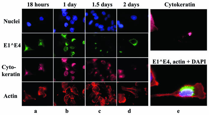
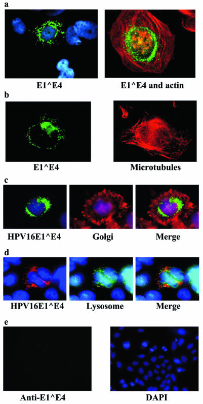
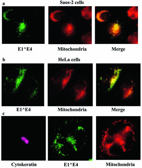
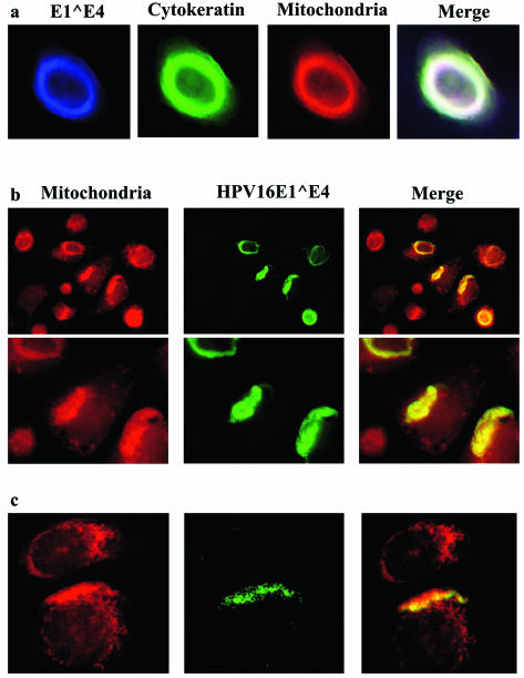
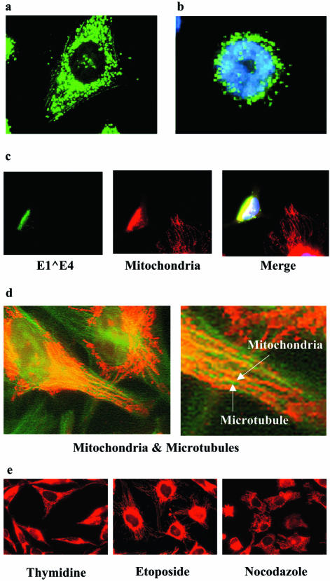
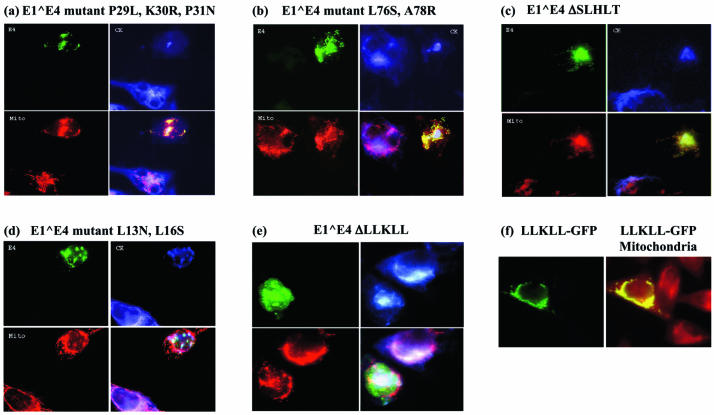
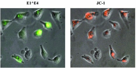
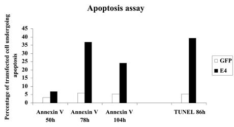
The human papillomavirus (HPV) E1 empty set E4 protein is the most abundantly expressed viral protein in HPV-infected epithelia. It possesses diverse activities, including the ability to bind to the cytokeratin network and to DEAD-box proteins, and in some cases induces the collapse of the former. E1 empty set E4 is also able to prevent the progression of cells into mitosis by arresting them in the G(2) phase of the cell cycle. In spite of these intriguing properties, the role of this protein in the life cycle of the virus is not clear. Here we report that after binding to and collapsing the cytokeratin network, the HPV type 16 E1 empty set E4 protein binds to mitochondria. When cytokeratin is not present in the cell, E1 empty set E4 appears associated with mitochondria soon after its synthesis. The leucine cluster within the N-terminal portion of the E1 empty set E4 protein is pivotal in mediating this association. After the initial binding to mitochondria, the E1 empty set E4 protein induces the detachment of mitochondria from microtubules, causing the organelles to form a single large cluster adjacent to the nucleus. This is followed by a severe reduction in the mitochondrial membrane potential and an induction of apoptosis. HPV DNA replication and virion production occur in terminally differentiating cells which are keratin-rich, rigid squamae that exfoliate after completion of the differentiation process. Perturbation of the cytokeratin network and the eventual induction of apoptotic properties are processes that could render these unyielding cells more fragile and ease the exit of newly synthesized HPVs for subsequent rounds of infection.
Figures
Similar articles
-
The E1E4 protein of human papillomavirus type 16 associates with a putative RNA helicase through sequences in its C terminus.J Virol. 2000 Nov;74(21):10081-95. doi: 10.1128/jvi.74.21.10081-10095.2000. J Virol. 2000. PMID: 11024137 Free PMC article.
-
The human papillomavirus type 11 E1E4 protein is phosphorylated in genital epithelium.Virology. 2000 Mar 15;268(2):430-9. doi: 10.1006/viro.1999.0173. Virology. 2000. PMID: 10704351
-
E1--E4-mediated keratin phosphorylation and ubiquitylation: a mechanism for keratin depletion in HPV16-infected epithelium.J Cell Sci. 2010 Aug 15;123(Pt 16):2810-22. doi: 10.1242/jcs.061978. Epub 2010 Jul 27. J Cell Sci. 2010. PMID: 20663917 Free PMC article.
-
Keratinocyte Differentiation-Dependent Human Papillomavirus Gene Regulation.Viruses. 2017 Aug 30;9(9):245. doi: 10.3390/v9090245. Viruses. 2017. PMID: 28867768 Free PMC article. Review.
-
Pathogenesis of human papillomaviruses in differentiating epithelia.Microbiol Mol Biol Rev. 2004 Jun;68(2):362-72. doi: 10.1128/MMBR.68.2.362-372.2004. Microbiol Mol Biol Rev. 2004. PMID: 15187189 Free PMC article. Review.
Cited by
-
Mitochondrial Proteins Coded by Human Tumor Viruses.Front Microbiol. 2018 Feb 6;9:81. doi: 10.3389/fmicb.2018.00081. eCollection 2018. Front Microbiol. 2018. PMID: 29467726 Free PMC article. Review.
-
Detection of human papillomavirus in cases of head and neck squamous cell carcinoma by RNA-seq and VirTect.Mol Oncol. 2019 Apr;13(4):829-839. doi: 10.1002/1878-0261.12435. Epub 2019 Feb 23. Mol Oncol. 2019. PMID: 30597724 Free PMC article.
-
Identification of an arginine-rich motif in human papillomavirus type 1 E1;E4 protein necessary for E4-mediated inhibition of cellular DNA synthesis in vitro and in cells.J Virol. 2008 Sep;82(18):9056-64. doi: 10.1128/JVI.01080-08. Epub 2008 Jul 16. J Virol. 2008. PMID: 18632869 Free PMC article.
-
AKT1 loss correlates with episomal HPV16 in vulval intraepithelial neoplasia.PLoS One. 2012;7(6):e38608. doi: 10.1371/journal.pone.0038608. Epub 2012 Jun 7. PLoS One. 2012. PMID: 22685591 Free PMC article.
-
Human papillomavirus 18 E1^E4 protein interacts with cyclin A/CDK 2 through an RXL motif.Mol Cell Biochem. 2013 Jan;373(1-2):29-40. doi: 10.1007/s11010-012-1472-y. Epub 2012 Oct 13. Mol Cell Biochem. 2013. PMID: 23065011
References
-
- Bryan, J. T., and D. R. Brown. 2000. Association of the human papillomavirus type 11 E1-E4 protein with cornified cell envelopes derived from infected genital epithelium. Virology 277:262-269. - PubMed
-
- Bryan, J. T., K. H. Fife, and D. R. Brown. 1998. The intracellular expression pattern of the human papillomavirus type 11 E1-E4 protein correlates with its ability to self associate. Virology 241:49-60. - PubMed
-
- Choo, K. B., C. C. Pan, and S. H. Han. 1987. Integration of human papillomavirus type 16 into cellular DNA of cervical carcinoma: preferential deletion of the E2 gene and invariable retention of the long control region and the E6/E7 open reading frames. Virology 161:259-261. - PubMed
Publication types
MeSH terms
Substances
Grants and funding
LinkOut - more resources
Full Text Sources
Other Literature Sources
