. 2004 Jul;78(13):7264-9.
doi: 10.1128/JVI.78.13.7264-7269.2004.

Intracellular approach for blocking JC virus gene expression by using RNA interference during viral infection

Affiliations

Intracellular approach for blocking JC virus gene expression by using RNA interference during viral infection

Sujatha Radhakrishnan et al. J Virol. 2004 Jul.

Abstract

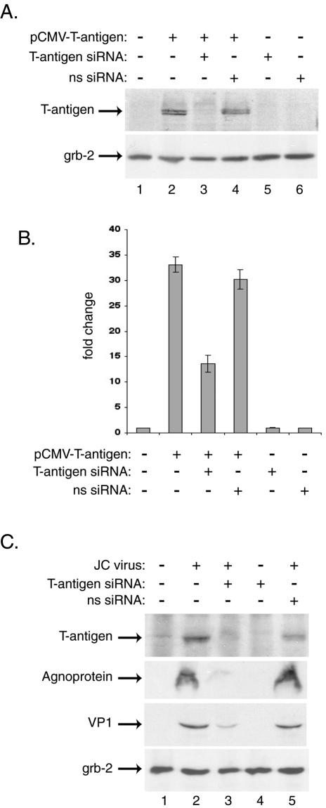
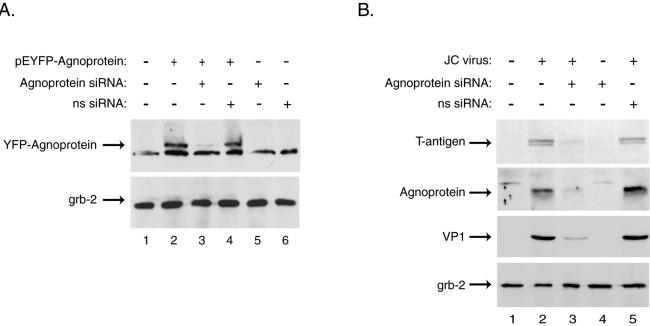
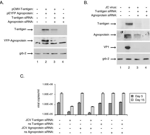
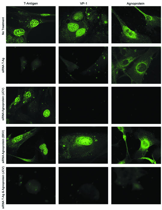
The human polyomavirus, JC virus (JCV), encodes two regulatory proteins at the early (T antigen) and the late (agnoprotein) phases of viral infection whose activities are important for the production of the viral capsid proteins and the dysregulation of several host factors and their functions. For this study, we designed and utilized an RNA interference strategy via small interfering RNAs (siRNAs) that targeted the expression of T antigen and agnoprotein in human astrocytic cells. The treatment of cells with specific siRNA oligonucleotides targeting a conserved region of T antigen, nucleotides (nt) 4256 to 4276 (Mad-1 strain), caused a >50% decline in the level of T antigen and in its transcriptional activity upon the viral capsid genes as well as a significant reduction in viral DNA replication in infected cells. Similarly, a single siRNA that aimed at nt 324 to 342 of agnoprotein noticeably reduced early and late viral protein production. A combined treatment of the infected cells with both T-antigen and agnoprotein siRNAs completely abolished viral capsid protein production, indicative of the ability of the siRNAs to effectively halt multiplication of the virus in infected cells. These observations provide a new avenue for possible treatments of patients with the JCV-induced demyelinating disease progressive multifocal leukoencephalopathy.


Figures

FIG. 1.
FIG. 1.
JCV T-antigen siRNA decreases expression of JCV proteins in transiently transfected and infected primary human astrocytes. Primary human fetal astrocytes were prepared as described previously and were seeded into six-well plates at a density of 500,000 cells/well (20). For transient transfections, cells were transfected by using FuGENE 6 with plasmid expressing JCV T antigen (15). The following day, the cells were transfected with double-stranded 21-bp siRNA for JCV T antigen targeting nt 4256 to 4276 of the Mad-1 isolate of JCV (sense strand, 5′-AAGUCUUUAGGGUCUUCUACCUdTdT-3′), while a nonspecific RNA (ns siRNA) targeted nt 4406 to 4426 of the reference strain 776 of SV40 (sense strand, 5′AAGUCCUUGGGGUCUUCUACCUdTdT-3′). The two base pair mismatches between the JCV and SV40 T antigens are underlined. The siRNAs were prepared as double-stranded, 2′-deprotected, and desalted oligonucleotides and were utilized according to the manufacturer's directions (Dharmacon). For the transfection of siRNAs, 100 pmol of siRNA was mixed with 3 μl of Oligofectamine (Invitrogen), diluted in OptiMEM (Invitrogen), and incubated with the cell cultures for 4 h at 37°C under serum- and antibiotic-free conditions. After transfection, the cells were fed with serum-containing medium without removing the siRNA transfection mixture. (A) Whole-cell extracts prepared from transfected astrocytes 24 h after siRNA treatment were analyzed by Western blotting for the presence of T antigen (pAb416; Oncogene Science) and the unrelated protein Grb-2 (upper and lower panels, respectively). (B) In parallel, samples transfected with 1.0 μg of JCV T-antigen expression plasmid along with 0.5 μg of a luciferase reporter construct containing the JCV late promoter (Mad-1 strain) were harvested 24 h after siRNA treatment, and luciferase activity was measured according to the manufacturer's directions (Promega luciferase assay system). Activities are presented as fold changes from the background activity of the JCV late promoter, arbitrarily set as 1. Data are means from four experiments, and standard deviations are indicated by error bars. (C) Primary astrocytes were infected with the JCV Mad-4 strain at a multiplicity of infection of 1 in serum-free medium for 3 h at 37°C. Uninfected and infected cells were then transfected with T-antigen siRNA at days 1, 5, and 10 postinfection and were harvested at day 15. Western blotting was performed on whole-cell extracts for the presence of JCV early and late proteins T antigen (pAb416; Oncogene Research Products), agnoprotein (7), and VP1 (pAb597; kindly provided by Walter Atwood, Brown University) as well as the cellular protein Grb-2 (pAb81; BD Biosciences). Proteins were visualized by using horseradish peroxidase-conjugated secondary antibodies and the ECL-Plus system (Amersham).
FIG. 2.
FIG. 2.
JCV agnoprotein siRNA decreases agnoprotein expression as well as that of other viral proteins in primary human astrocytes. Primary human fetal astrocyte preparations, transient transfections, siRNA treatments, and Western blotting were performed as described in the text and the legend to Fig. 1. The cells were transfected with a plasmid containing JCV agnoprotein fused to YFP (6). The JCV agnoprotein siRNA targeted nt 324 to 342 of the Mad-1 isolate of JCV (sense strand, 5′-AACCUGGAGUGGAACUAAAdTdT-3′), while a nonspecific siRNA (ns siRNA) targeted nt 435 to 453 of the Dunlop strain of BKV (sense strand, 5′-AACCUGGACUGGAACAAAAdTdT-3′). The two base pair mismatches between JCV and BKV agnoprotein sequences are underlined. (A) Whole-cell extracts prepared from transfected astrocytes 24 h after treatment with specific or nonspecific siRNA were analyzed by Western blotting for the presence of agnoprotein and the unrelated cellular factor Grb-2 (upper and lower panels, respectively). (B) Primary astrocytes that were uninfected or infected with the JCV Mad-4 strain were then transfected with JCV agnoprotein or nonspecific BKV agnoprotein siRNA at days 1, 5, and 10 postinfection and were harvested at day 15. Western blotting was performed on whole-cell extracts for presence of the JCV T antigen, agnoprotein, and VP1 as well as the cellular protein Grb-2.
FIG. 3.
FIG. 3.
Treatment with siRNAs targeting JCV T antigen and agnoprotein abolishes their expression, as well as that of the JCV late protein VP1, in primary human astrocytes and affects viral replication in infected cells. Primary human fetal astrocyte preparations, transient transfections, siRNA treatments, and Western blotting were performed as described in the text and the legend to Fig. 1. (A) Whole-cell extracts from astrocytes transfected with expression plasmids for JCV T antigen, YFP-agnoprotein, or both were prepared from cultures 24 h after siRNA treatment and were analyzed by Western blotting for the presence of T antigen and agnoprotein as well as for the unrelated protein Grb-2. (B) Astrocyte cultures were infected with the JCV Mad-4 strain and were then transfected with siRNAs targeting JCV T antigen, agnoprotein, or both at days 1, 5, and 10 postinfection. Western blotting was performed on whole-cell extracts harvested at day 15 postinfection for the presence of JCV T antigen and agnoprotein as well as the viral late protein, VP1, and the cellular protein Grb-2. Supernatants collected from infected and siRNA-treated cells at days 5 and 15 postinfection were analyzed by quantitative real-time PCR for the presence of replicated JCV DNA essentially as described previously (17). The PCRs included JCV-specific forward and reverse primers (200 and 400 nM) representing nt 2393 to 2412 and 2468 to 2486 of the Mad-1 strain of JCV plus 200 nM JCV-specific probe (nt 2428 to 2458) fluorescently labeled at the 5′ and 3′ ends with FAM and BHQ1, respectively. Five microliters of cell culture supernatant was directly analyzed in triplicate in 50-μl reaction mixtures containing the above primers and probe in 1× TaqMan Universal Master Mix (Perkin-Elmer). Plasmid DNA containing the JCV genome was used to generate a standard curve against which the samples were analyzed using iCycler software (Bio-Rad).
FIG. 4.
FIG. 4.
Immunocytochemistry of primary astrocytes infected with JCV reveals alterations in JCV protein levels upon JCV T-antigen and agnoprotein siRNA treatment. Cells were infected with JCV and treated at days 1, 5, and 10 postinfection with JCV T-antigen siRNA, JCV agnoprotein siRNA, or both. The cells were also treated with nonspecific agnoprotein siRNA. The cells were subcultured and plated onto poly-l-lysine-coated chamber slides (Falcon) on day 13 and were fixed on day 15 postinfection with ice-cold acetone for 3 min. Viral proteins were detected by immunocytochemistry as described previously (20), using the same primary antibodies as for the Western blot analysis (see the legend to Fig. 1). The proteins were visualized with fluorescein-conjugated secondary antibodies.

Similar articles

Cited by

References

    1. Aksamit, A. 2001. Treatment of non-AIDS progressive multifocal leukoencephalopathy with cytosine arabinoside. J. Neurovirol. 7:386-390. - PubMed
    1. Berger, J. R., and M. Concha. 1995. Progressive multifocal leukoencephalopathy: the evolution of a disease once considered rare. J. Neurovirol. 1:5-18. - PubMed
    1. Chang, J., and J. M. Taylor. 2003. Susceptibility of human hepatitis delta virus RNAs to small interfering RNA action. J. Virol. 77:9728-9731. - PMC - PubMed
    1. Cinque, P., I. J. Koralnik, and D. B. Clifford. 2003. The evolving face of human immunodeficiency virus-related progressive multifocal leukoencephalopathy: defining a consensus terminology. J. Neurovirol. 9(Suppl. 1):88-92. - PubMed
    1. Clifford, D. B., and E. O. Major. 2001. The biology of JC virus and progressive multifocal leukoencephalopathy. J. Neurovirol. 4:279. - PubMed

Publication types

MeSH terms

Substances