

doi: 10.1128/JVI.78.13.7270-7273.2004.
Inhibition of virus production in JC virus-infected cells by postinfection RNA interference
Affiliations
- PMID: 15194803
- PMCID: PMC421637
- DOI: 10.1128/JVI.78.13.7270-7273.2004
Item in Clipboard
Inhibition of virus production in JC virus-infected cells by postinfection RNA interference
J Virol.
2004 Jul.
Abstract
RNA interference has been applied for the prevention of virus infections in mammalian cells but has not succeeded in eliminating infections from already infected cells. We now show that the transfection of JC virus-infected SVG-A human glial cells with small interfering RNAs that target late viral proteins, including agnoprotein and VP1, results in a marked inhibition both of viral protein expression and of virus production. RNA interference directed against JC virus genes may thus provide a basis for the development of new strategies to control infections with this polyomavirus.
Figures
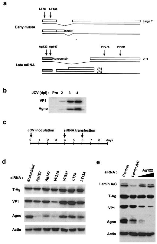
Effects of postinfection RNAi on the abundance of JCV proteins in JCV-infected SVG-A cells. (a) Schematic representation of major early and late mRNAs of JCV. Major early mRNAs encode the small t antigen and T-Ag, which are translated as splicing variants. Two major forms of late mRNA encode either agnoprotein and VP1 or agnoprotein, VP2, and VP3. The regions of the viral RNAs targeted by the siRNAs are indicated by arrows. (b) Immunoblot analysis of the abundance of VP1 and agnoprotein of JCV in SVG-A cells at the indicated times after infection with JCV. (c) Schedule for JCV infection and siRNA transfection in SVG-A cells. (d) Immunoblot analysis of the indicated proteins in JCV-infected cells subjected to transfection with the indicated siRNAs. (e) Immunoblot analysis of the indicated proteins in JCV-infected cells subjected to transfection with the Ag122 siRNA at 60 or 120 pmol/well or with a lamin A/C-specific siRNA. Control (infected) cells were subjected to mock transfection.
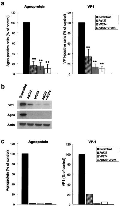
Indirect immunofluorescence analysis of the effects of RNAi on viral protein expression in JCV-infected SVG-A cells. (a) The proportion of cells that were positive for agnoprotein or VP1 was determined by an indirect immunofluorescence analysis 48 h after transfection with the indicated siRNA(s). The data are expressed as percentages of the proportion determined for JCV-infected cells transfected with the scrambled siRNA (control) and are means ± standard deviations (SD) of values from at least three independent experiments. **, P < 0.02 versus the value for cells transfected with the scrambled siRNA (Student's t test). (b) Immunoblot analysis of VP1, agnoprotein, and actin expression in JCV-infected cells transfected with the indicated siRNA(s). (c) The signals for agnoprotein and VP1 were quantified with an image analyzer and expressed as percentages of the value for cells transfected with the scrambled siRNA.
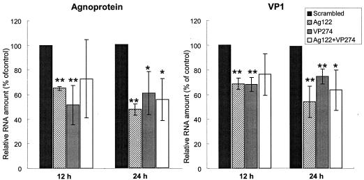
Depletion of viral RNAs by RNAi in JCV-infected SVG-A cells. Total RNAs isolated from JCV-infected cells 12 or 24 h after transfection with the indicated siRNAs were subjected to an RT-PCR analysis of agnoprotein and VP1 mRNAs. The data were normalized to the amount of β-actin mRNA and are expressed as percentages of the normalized value for JCV-infected cells transfected with the scrambled siRNA (control); they are means ± SD of values from at least three independent experiments. *, P < 0.05, and **, P < 0.02 versus the value for cells transfected with the scrambled siRNA.
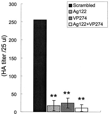
Inhibition of JCV production by RNAi in SVG-A cells. Extracts prepared from JCV-infected cells 36 h after transfection with the indicated siRNAs were assayed for hemagglutination activity (HA). The data are expressed as HA titers per 25 μl of cell extract and are means ± SD of values from at least three independent experiments. **, P < 0.02 versus the value for cells transfected with the scrambled siRNA.
Similar articles
-
Intracellular approach for blocking JC virus gene expression by using RNA interference during viral infection.J Virol. 2004 Jul;78(13):7264-9. doi: 10.1128/JVI.78.13.7264-7269.2004. J Virol. 2004. PMID: 15194802 Free PMC article.
-
[Recent research on the JC virus].No To Shinkei. 2007 Feb;59(2):101-8. No To Shinkei. 2007. PMID: 17315751 Review. Japanese.
-
Functional interaction between JC virus late regulatory agnoprotein and cellular Y-box binding transcription factor, YB-1.J Virol. 2002 Apr;76(8):3828-38. doi: 10.1128/jvi.76.8.3828-3838.2002. J Virol. 2002. PMID: 11907223 Free PMC article.
-
Role of JC virus agnoprotein in virion formation.Microbiol Immunol. 2012 Sep;56(9):639-46. doi: 10.1111/j.1348-0421.2012.00484.x. Microbiol Immunol. 2012. PMID: 22708997
-
[Recent research on the JC virus].Brain Nerve. 2007 Feb;59(2):101-8. Brain Nerve. 2007. PMID: 17380774 Review. Japanese.
Cited by
-
Progressive multifocal leukoencephalopathy: current treatment options and future perspectives.Ther Adv Neurol Disord. 2015 Nov;8(6):255-73. doi: 10.1177/1756285615602832. Ther Adv Neurol Disord. 2015. PMID: 26600871 Free PMC article. Review.
-
Progressive multifocal leukoencephalopathy: clinical and molecular aspects.Rev Med Virol. 2012 Jan;22(1):18-32. doi: 10.1002/rmv.710. Epub 2011 Sep 21. Rev Med Virol. 2012. PMID: 21936015 Free PMC article. Review.
-
Dissociation of heterochromatin protein 1 from lamin B receptor induced by human polyomavirus agnoprotein: role in nuclear egress of viral particles.EMBO Rep. 2005 May;6(5):452-7. doi: 10.1038/sj.embor.7400406. EMBO Rep. 2005. PMID: 15864296 Free PMC article.
-
Avian metapneumovirus phosphoprotein targeted RNA interference silences the expression of viral proteins and inhibits virus replication.Antiviral Res. 2006 Jan;69(1):46-51. doi: 10.1016/j.antiviral.2005.09.004. Epub 2005 Nov 15. Antiviral Res. 2006. PMID: 16310868 Free PMC article.
-
Regulation of gene expression in primate polyomaviruses.J Virol. 2009 Nov;83(21):10846-56. doi: 10.1128/JVI.00542-09. Epub 2009 Jul 29. J Virol. 2009. PMID: 19640999 Free PMC article. Review.
References
-
- Andino, R. 2003. RNAi puts a lid on virus replication. Nat. Biotechnol. 21:629-630. - PubMed
-
- Clifford, D. B. 1999. Opportunistic viral infections in the setting of human immunodeficiency virus. Semin. Neurol. 19:185-192. - PubMed
-
- Endo, S., Y. Okada, Y. Orba, H. Nishihara, S. Tanaka, K. Nagashima, and H. Sawa. 2003. JC virus agnoprotein colocalizes with tubulin. J. Neurovirol. 9(Suppl. 1):10-14. - PubMed
Publication types
MeSH terms
Substances
LinkOut - more resources
Full Text Sources
Other Literature Sources
