

Influence of N-glycans on processing and biological activity of the nipah virus fusion protein
- PMID: 15194804
- PMCID: PMC421684
- DOI: 10.1128/JVI.78.13.7274-7278.2004
Influence of N-glycans on processing and biological activity of the nipah virus fusion protein
Abstract
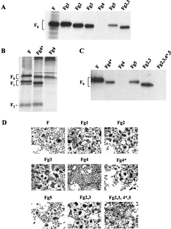
Nipah virus (NiV), a new member of the Paramyxoviridae, codes for a fusion (F) protein with five potential N-glycosylation sites. Because glycans are known to be important structural components affecting the conformation and function of viral glycoproteins, we analyzed the effect of the deletion of N-linked oligosaccharides on cell surface transport, proteolytic cleavage, and the biological activity of the NiV F protein. Each of the five potential glycosylation sites was removed either individually or in combination, revealing that four sites are actually utilized (g2 and g3 in the F(2) subunit and g4 and g5 in the F(1) subunit). While the removal of g2 and/or g3 had no or little effect on cleavage, surface transport, and fusion activity, the elimination of g4 or g5 reduced the surface expression by more than 80%. Similar to a mutant lacking all N-glycans, g4 deletion mutants in which the potential glycosylation site was destroyed by introducing a glycine residue were neither cleaved nor transported to the cell surface and consequently were not able to mediate cell-to-cell fusion. This finding indicates that in the absence of g4, the amino acid sequence around position 414 is important for folding and transport.
Figures


Similar articles
-
Ubiquitous activation of the Nipah virus fusion protein does not require a basic amino acid at the cleavage site.J Virol. 2004 Sep;78(18):9705-12. doi: 10.1128/JVI.78.18.9705-9712.2004. J Virol. 2004. PMID: 15331703 Free PMC article.
-
N-glycans on Nipah virus fusion protein protect against neutralization but reduce membrane fusion and viral entry.J Virol. 2006 May;80(10):4878-89. doi: 10.1128/JVI.80.10.4878-4889.2006. J Virol. 2006. PMID: 16641279 Free PMC article.
-
Influence of N-linked oligosaccharide chains on the processing, cell surface expression and function of the measles virus fusion protein.J Gen Virol. 1995 Mar;76 ( Pt 3):705-10. doi: 10.1099/0022-1317-76-3-705. J Gen Virol. 1995. PMID: 7897359
-
Functional analysis of the N-linked glycans within the fusion protein of respiratory syncytial virus.Methods Mol Biol. 2007;379:69-83. doi: 10.1007/978-1-59745-393-6_5. Methods Mol Biol. 2007. PMID: 17502671 Review.
-
Virus activation by host proteinases. A pivotal role in the spread of infection, tissue tropism and pathogenicity.Microbiol Immunol. 1995;39(1):1-9. doi: 10.1111/j.1348-0421.1995.tb02161.x. Microbiol Immunol. 1995. PMID: 7783672 Review. No abstract available.
Cited by
-
Canine distemper viruses expressing a hemagglutinin without N-glycans lose virulence but retain immunosuppression.J Virol. 2010 Mar;84(6):2753-61. doi: 10.1128/JVI.01813-09. Epub 2009 Dec 30. J Virol. 2010. PMID: 20042514 Free PMC article.
-
Co-assembly of viral envelope glycoproteins regulates their polarized sorting in neurons.PLoS Pathog. 2014 May 15;10(5):e1004107. doi: 10.1371/journal.ppat.1004107. eCollection 2014 May. PLoS Pathog. 2014. PMID: 24831812 Free PMC article.
-
Activation of the Nipah virus fusion protein in MDCK cells is mediated by cathepsin B within the endosome-recycling compartment.J Virol. 2012 Apr;86(7):3736-45. doi: 10.1128/JVI.06628-11. Epub 2012 Jan 25. J Virol. 2012. PMID: 22278224 Free PMC article.
-
Biochemical, conformational, and immunogenic analysis of soluble trimeric forms of henipavirus fusion glycoproteins.J Virol. 2012 Nov;86(21):11457-71. doi: 10.1128/JVI.01318-12. Epub 2012 Aug 22. J Virol. 2012. PMID: 22915804 Free PMC article.
-
Endocytosis of the Nipah virus glycoproteins.J Virol. 2005 Mar;79(6):3865-72. doi: 10.1128/JVI.79.6.3865-3872.2005. J Virol. 2005. PMID: 15731282 Free PMC article.
References
-
- Bagai, S., and R. A. Lamb. 1995. Individual roles of N-linked oligosaccharide chains in intracellular transport of the paramyxovirus SV5 fusion protein. Virology 209:250-256. - PubMed
-
- Chen, L., J. J. Gorman, J. McKimm-Breschkin, L. J. Lawrence, P. A. Tulloch, B. J. Smith, P. M. Colman, and M. C. Lawrence. 2001. The structure of the fusion glycoprotein of Newcastle disease virus suggests a novel paradigm for the molecular mechanism of membrane fusion. Structure 9:255-266. - PubMed
-
- Doms, R. W., R. A. Lamb, J. K. Rose, and A. Helenius. 1993. Folding and assembly of viral membrane proteins. Virology 193:545-562. - PubMed
-
- Hooper, P., S. Zaki, P. Daniels, and D. Middleton. 2002. Comparative pathology of the diseases caused by Hendra and Nipah viruses. Microbes Infect. 3:315-322. - PubMed
Publication types
MeSH terms
Substances
LinkOut - more resources
Full Text Sources
Miscellaneous
